No pain, no gain : double network hydrogels get stronger after mechanical loading

Original paper: Matsuda et al., Mechanoresponsive self-growing hydrogels inspired by muscle training, Science 363, 504-508 (2019)


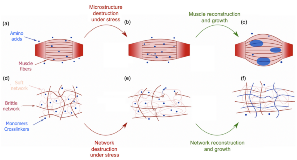
During the COVID-19 lockdown, many of us had the opportunity to workout, for instance by lifting weights to build stronger biceps. During a workout, our muscles undergo some damage at the micrometer scale that triggers an immune system response (Figure 1a). Adequate amino acids, the main constituents of proteins that give muscles their structure, are then carried close to the torn tissues to repair the damage and grow new muscle thanks to the binding between the amino acids and the tissues [1]. This process is a sort of mechanical solicitation where your body creates stronger biological materials with more mass (Figure 1b-c). This seems, however, counter-intuitive when compared to the response of synthetic soft materials under the same mechanical solicitation. Indeed, mechanical stress usually weakens or even damages synthetic materials. For instance, pulling too hard on an elastic band will result in its irreversible failure. Can we then use muscle growth as an inspiration to design materials that would get stronger and bigger under mechanical stress? By mimicking this process, a group of scientists from Hokkaido University developed such a material. This new material belongs to the family of polymer hydrogels. Hydrogels consist of a stretchable 3D network of polymer chains in water connected to each other by molecules called crosslinkers. These crosslinkers control the stiffness of the network. Indeed, a highly connected network will require more stress to be deformed than a loosely connected one.

The difference is that this new material is a double network hydrogel  — a hydrogel with two interpenetrated networks — one of them is brittle and rigid due to a high content of crosslinkers, shown as red chains in Figure 1d, while the other one is soft and stretchable due to low content of crosslinkers, shown as pink chains in Figure 1d. This double network is immersed in a solution containing 80-90% water and two types of molecules that are the building blocks of the hydrogel: monomers and crosslinkers. When a tensile stress is applied to the material, the brittle network is the one that “feels” the pressure, leading to breakage of its strands while leaving the soft network undamaged (Figure 1e). During the failure of those polymer chains, the bonds between the carbon atoms break down due to the stress which generates highly reactive chemical species known as radicals [2]. These radicals initiate the formation of new polymer chains by reacting with the building blocks in the solution [3]. As these new chains are also connected by crosslinkers during the reaction, the damaged network is then not only restored but extended and strengthened [4].

Figure 1. Illustration of the bio-inspired study on strengthening double network hydrogels under mechanical stress. Top: a muscle initially at rest (a), after being damaged under effort (b) and reconstructed and grown by the action of neighboring amino acids (c). Bottom: a double network hydrogel at rest (d), after being damaged by tensile stress (e) and reconstructed and grown by the action of neighboring building monomers and crosslinkers (f)

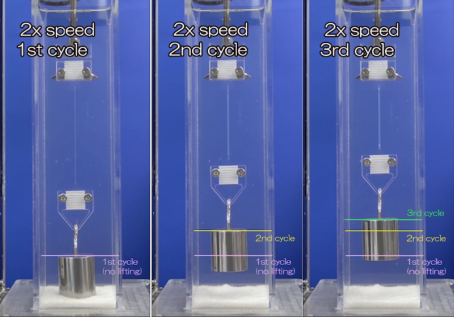
The most remarkable result which validates this process is shown in the following movie :

Movie 1. Cyclic test of tension and recovery of a double network hydrogel caught between two clamps. The setup is immersed in a solution of monomers and crosslinkers suitable for reconstruction and growth of the material after mechanical solicitation.

A double network hydrogel is stretched at constant stress three times in a row. The hydrogel is immersed in a solution of monomers and crosslinkers that will be used up for the brittle network reconstruction. After each cycle of stretching, a one-hour reconstruction and growth period allows the material to strengthen. Thanks to that stretching and recovering protocol, the material is able to lift a 200g weight higher and higher as it gets stronger.

The successful emulation of muscle self-strengthening applied to synthetic materials design paves the way for tremendous new bio-inspired materials. They are of especially great interest in soft robotics, where soft materials are exposed to harsh conditions that may lead to damage, like cuts. Rather than undergoing simple regeneration, the material would respond to damage by strengthening to better resist its environment.

[1] https://www.youtube.com/watch?v=2tM1LFFxeKg

[2] Polymer Mechanochemistry: Manufacturing Is Now a Force to Be Reckoned With

[3] This polymerization reaction is called radical polymerization. The radicals, R, in Figure 1e will react with the monomers which contain chemical groups sensitive to radicals (double carbon bonds for instance). The result of this first reaction is another radical which will react with another monomer, thus, by repeating the process, connecting the monomers into a polymer chain.

[4] The generation of enough radicals and their contact with monomers and crosslinkers are assured by the second soft network which maintains the cohesion of the whole material while bonds are broken. This is comparable to a muscle where the breakage of its microstructure does not provoke the collapse of the entire muscle.

Who needs polymer physics when you can get worms drunk instead?

Original paper: Rheology of Entangled Active Polymer-Like T. Tubifex Worms (arXiv here)


If you speak to a soft matter physicist these days, within a few minutes the term “active matter” is bound to come up. A material is considered “active” when it burns energy to produce work, just like all sorts of molecular motors, proteins, and enzymes do inside your body. In this study, the scientists are focusing specifically on active polymers. These are long molecules which can burn energy to do physical work. Much of biological active matter is in the form of polymers (DNA or actin-myosin systems for example), and understanding them better would give direct insight into biophysics of all kinds. But polymers are microscopic objects with complex interactions, making them difficult to manipulate directly. To make matters worse, physicists have yet to fundamentally understand the behaviors of active materials, since they do not fit into our existing theories of so-called “passive” systems. In this study, Deblais and colleagues decided to entirely circumvent this problem by working with a much larger and easier-to-study system that behaves similarly to a polymer solution: a mixture of squirming worms in water.

The researchers focused on the viscous properties of this living material, which behaves somewhat like a fluid. Viscosity is a measure of a fluid’s resistance to gradients in the flow. Polymer fluids are highly viscous because the long molecules in a polymeric liquid get tangled up in one another. Physical descriptions of most fluids assume that viscosity is a constant (so called Newtonian fluids), but many materials exhibit what is called shear thinning. This is when a fluid flows more easily as one applies an increasing shear force, that is, a force pulling the system apart. We encounter shear thinning at the dinner table all the time when struggling to pour ketchup, another polymeric fluid, out of a bottle. If the bottle is shaken fast enough, increasing the shear force applied, the ketchup flows smoothly like a liquid. In polymer systems (like xanthan gum in the ketchup) shear thinning happens when polymers are pulled apart fast enough that they tend to align together, which loosens the entanglements that held the system together before. 

In this study, the researchers asked: how does shear thinning behavior change if the polymers in question were alive? To answer this question, they set out to measure the shear thinning properties of a mixture of worms at various levels of worm activity. Here, “worm activity” refers to how fast the worm is wriggling, which is calculated by measuring how quickly the distance between the two ends of a given worm changes. This leads to two logistical questions: how is the level of worm activity being modified, and how is the viscosity being measured?

Figure 1. This movie shows two worms, one in water (left) and one in a water + alcohol mixture (right). The worm on the right shows a decrease in activity when they are exposed to alcohol, which is one of the two ways the researchers modified worm activity in this study. Video taken from the original article.

The answer to the first question should be familiar to many humans. To make the worms less active, they were put into a solution containing water and a small amount of ethanol, the same type of alcohol found in beer, wine, and spirits. Once the worms were nice and drunk, the researchers noticed that they squirmed about more slowly, as shown in Figure 1. Thankfully, when the ethanol was removed, the worms returned to their previous level of activity! To make sure the alcohol wasn’t doing anything funny to the worms, they found a second way to reduce the activity — by reducing the temperature of the worm solution. Colder temperatures made for more chilled out worms, no pun intended.

Figure 2. This movie shows the functioning of the rheometer. The worms are placed inside a chamber between two plates. The top plate rotates with respect to the bottom plate, and the response of the material is measured. Video taken from the original article.

The researchers used a device called a parallel-plate rheometer to understand the shear thinning behavior of this living polymer system. As seen in Figure 2, a parallel-plate rheometer sandwiches a sample in between two flat plates and viscosity is measured by determining how much force is necessary to rotate the top plate, effectively pushing the material by twisting its surface. The viscosity of the worm mixtures was first determined at three different temperatures, and for worms drunk on ethanol. The results were surprising! The rheological behaviour of the low-activity worm mixtures matched with theories of polymer shear thinning quite well. It seems the worms have the same alignment properties as passive polymer solutions under shear!

So what happens when the worms are sober, more active, and wriggling around? They saw that the required twisting rate needed to thin the mixture decreased. In this case, the worm activity allowed for easier and quicker rearrangement while the mixture was pulled apart by the rheometer’s twisting motion. One can imagine that instead of needing to pull all the worms to the point of alignment, it may have been enough to nudge them in that direction and their wriggling did the rest. We can now imagine that the same thing might be true for non-living polymers: if a polymer material with shear thinning behavior is given an extra source of activity, then its thinning behavior may become more significant. 

The lesson to be learned here is partly about worms, polymers, and the adverse effects of ethanol, but really this experiment is a testament to the power and generality of physical descriptions. This study teaches us about the possible behavior of an active polymer system with processes that are relevant on the scale of a few micrometers, by studying real life worms that you can see with the naked eye! In general, it is usually possible to find analog systems that have the desired properties for your study, but which are easier to manipulate. Physics then gives you the bridge between the system of interest and your simpler analog, allowing you to harness the power of interdisciplinary science to ask questions previously unanswerable.

Featured image for the article is taken from the original article.

Supersoft matter bounces back: Softer, Better, Stronger

Original paper: Solvent-free, supersoft and superelastic bottlebrush melts and networks


Polymers are made of long molecules (polymer chains) consisting of shorter, repeating units called monomers. Like cooked spaghetti noodles, many polymer chains coexist in the same shared space and when too many of them overlap entanglement may occur (Figure 1). Such entangled messes of polymer chains are stiff and hard to deform, limiting the elasticity of polymer-based synthetic materials. One way of softening materials is by disentangling the polymer chains via soaking the polymer chains in a solvent, such as water. The solvent molecules in hydrogels occupy space between polymer chains driving the chains away from each other, similar to how pouring water overcooked spaghetti drives the noodles apart. This led to the discovery of hydrogels, the primary component of soft contact lenses and tissue implants [1]. But if you’ve ever worn soft contact lenses, you may know that they dry out and harden if they are not stored in a solution. This pervasive issue of hydrogel materials occurs when the solvent leaks or evaporates, affecting their mechanical properties. In this week’s post, polymer scientists develop super-soft dry elastomers (very elastic or rubbery polymers) that surpass the softness and elasticity of hydrogels, all without getting their hands wet.

Figure 1. Spaghetti pomodoro e basilico. The noodles demonstrate how long, flexible objects intertwine with each other to form an entangled complex, resembling polymer networks in hydrogels. Image courtesy of Wikipedia.

What does it take to design a polymer material that intrinsically avoids entanglement without using a large amount of solvent? William Daniel and his colleagues tackle this issue by designing a polymer chain geometry resembling a bottlebrush (shown in Figure 2). The bottlebrush geometry consists of a linear polymer backbone onto which short side chains, called bristles, are grafted. These bottlebrush-shaped chains are soft even in the absence of solvent. Instead of relying on small solvent molecules, bottlebrush networks use their bristles to keep polymer chains away from each other because bristles are too short to participate in the entanglement. As a result, the bottlebrush chains have an overall repulsion effect that resembles the repulsion effect of solvent molecules in hydrogels. Bristle repulsion allows bottlebrush polymers to surpass the elasticity of hydrogels! As an example, Figure 3 shows a compression test where a bottlebrush elastomer (on the right) retains its structure whereas a hydrogel material (on the left) fractures when compressed. Despite their similar elastic moduli, the bottlebrush elastomer displayed much greater compressibility than the hydrogel.

Figure 2. Schematic of three bottlebrush polymer chains. Each bottlebrush chain consists of a polymer backbone (linear chain) onto which short side chains (bristles) are attached. Figure adapted from the original article.

But what is an elastic modulus and why does it matter? The elastic modulus is a measure of the stiffness of a material and is given by the ratio of stress, the force causing deformation per area, to strain, the relative length by which the material is deformed by the stress. In these terms, a small modulus corresponds to low force per area resulting in significant deformation during compression – exactly what we expect of soft materials! As the entanglement density increases, a polymer chain network becomes more crowded resulting in a stiffer material [2]. Since bottlebrush bristles are too short to entangle, increasing the bristle density further reduces the entanglement density. Thus the elastic modulus of bottlebrush elastomers can be tuned by controlling the number of bristles grafted onto the polymer backbone. These bristles comprise the majority of the mass of the elastomer, e.g. 87% of the mass of the elastomer described in Figure 3. 

Figure 3. Compression test of a hydrogel versus a bottlebrush elastomer. Left: PAM or poly(acrylamide) hydrogel made of 10% by weight polymer chains and 90% solvent. Right: bottlebrush elastomer made of 8% by weight backbone chains, 87% bristles and 5% solvent. Both materials have similar moduli (~2000 Pa). During compression, the bottlebrush elastomer kept its form while the hydrogel fractured. Figure adapted from the original article.

In addition to super-softness, bottlebrush networks are also highly compressible. The stress measurement in Figure 4 shows that bottlebrush elastomers (red curve) tolerated five times more stress before fracture compared to hydrogels (blue curve). Furthermore, the compression ratio (equilibrium length to compression length) of bottlebrushes was three times higher before fracturing. This means that bottlebrush elastomers are capable of sustaining much more deformation, and hence strain, than hydrogels. 

Figure 4. Stress measured during compression of a bottlebrush elastomer (red curve) and a hydrogel (blue curve). The bottlebrush elastomer achieved a compression ratio (equilibrium length to compressed length) of about 10 while the hydrogel fractured at a compression ratio equal to 3. Image adapted from the original paper.

The idea of attaching bristles onto a polymer backbone in high density gave William Daniel and his colleagues control over the stiffness due to entanglement. This work expands scientists’ understanding of material properties consisting of branched polymer chains and points to a new frontier of dry supersoft materials. These new materials could play an important role in the development of soft robotics and synthetic biological tissues.

[1] Swell gels (2002)

[2] In polymers, the entanglement contribution to the elastic modulus is given by the modulus equation:

Ge = neRT, 

where ne is the number of chains involved in entanglement per unit volume, R is the universal gas constant, and T is the temperature. The modulus equation suggests that polymer materials become stiffer when heated. As temperature increases, the polymer network gains kinetic energy to attain structures with more “randomness”, or entropy. This increased randomness in polymer networks leads to more entangled states (higher entropy) that makes the material less prone to lengthening, hence more resistant to deformation.

Lifehack: How to pack two meters of chromatin into your cell’s nucleus, knot-free!

Original paper: The fractal globule as a model of chromatin architecture in the cell


The entirety of our genetic information is encoded in our DNA. In our cells, it wraps together with proteins to form a flexible fiber about 2 metres long known as chromatin. Despite its length, each cell in our body keeps a copy of our chromatin in its nucleus, which is only about 10 microns across. For scale, if the nucleus was the size of a basketball, its chromatin would be  about 90 miles long. How can it all fit in there? To make matters worse, the cell needs chromatin to be easily accessible for reading and copying, so it can’t be all tangled up. It’s not surprising then that scientists have been puzzled as to how this packing problem can be reliably solved in every cell. The solution is to pack the chromatin in a specific way, and research suggests that this may be in the form of a “fractal globule”. 

An equilibrium globule is the state that a polymer (a long repetitive molecular sequence, like chromatin) takes when it is left for a long time in a liquid that doesn’t dissolve it well. In such a liquid, the polymer is more attracted to itself than the molecules around it, so it collapses into a globule to minimize the amount of contact between itself and its surroundings. The resulting object is much denser than typical polymers in good solvents and is dense enough to fit inside a nucleus. However, like stuffing headphone cables into your pocket, it develops many knots and its different regions mix with one another.

On the other hand, if you change the polymer’s environment fast enough that it doesn’t have the time to fully equilibrate, then every piece of the polymer will locally collapse into its own globule. In other words, the polymer forms a globule made of smaller globules and is called a fractal globule. Fractals are objects which look the same at all scales, like the edge of a cloud or the coastline of England. If you zoom in or out on either of these objects, they look more or less the same. This isn’t an “equilibrium” state, meaning it will slowly fall out of this configuration. However, until the whole polymer equilibrates (which takes a long time), the chain has many desirable properties.

Figure 1. Simulated examples of fractal (A,C) and equilibrium globules (B,D), showing compartmentalization of different portions of the polymer. The chain color goes from red to blue as shown above. Compartmentalization means that parts of the chromatin stay near other parts with the same color (adapted from paper [1]).

We are interested in these globule states because they are dense enough that a globule of chromatin can fit inside of a cell nucleus. But it’s not enough to simply fit inside; the cell needs chromatin to avoid forming knots, since getting tangled would prevent the cell from properly reading its own DNA. Live cells also keep their chromatin nicely compartmentalized, that is, different regions along the genome stay spatially separated from one another. Unlike equilibrium globules, fractal globules have few knots and are also compartmentalized! To get a better picture for what this means, Leonid Mirny performed simulations of the different types of globules. Figure 1 shows the results of these simulations, highlighting how different the two states look in terms of knotting and separation of regions of the polymer. 

So it seems that the fractal globule state has all the properties we need for a good model of chromatin! But, as scientists, we know that no matter how well a theory fits the characteristics we want it to have, we need experimental evidence before believing anything. In the case of this fractal globule model for genome organization, evidence has come in the form of “contact probability maps”. These are collected from large populations of cells whose DNA is cut, spliced, and read in such a way that allows for a measurement of the probability that any two sites on the chromatin are touching at any given time. Among other things, these maps give us information about how chromatin is packed. So the question becomes, “what does the fractal globule model predict a contact probability map to look like?”

The fractal globule model doesn’t make exact predictions about where one will find specific segments of chromatin, but it does predict a contact probability as a function of distance between two sites, s. Specifically, the model predicts that the contact probability between two sites scales like 1/s. Meaning, if I look at sites that are twice as far apart along the polymer, then they are half as likely to be touching. This 1/s scaling is what was observed on intermediate scales (about 100,000 to 6 million base pairs) by looking at contact probability maps averaged over a whole population of cells.

We still don’t know how the cell maintains and tunes this fractal globule state, and we still have not developed a dynamic version of this picture, which is necessary since it is well-established that the chromatin in our cells is far from static. But this study gives us a new picture of how chromatin is organized inside cells. It isn’t randomly configured like headphone cables in your pocket or a ball of yarn. Rather it is folded onto itself in a self-similar way. This model is attractively simple, requires little fine-tuning, all while producing a long-lived state with segregated territories and easily accessible genes. 

[1] Mirny, Leonid (2011), The fractal globule as a model of chromatin architecture in the cell. Chromosome Res.

Featured image for the article is taken from Wikimedia Commons.

How to turn off cancer cell growth using mechanosensing

Original paper: Stopping transformed cancer cell growth by rigidity sensing


Our bodies are made up of cells that can sense and respond to their dynamic environment. As an example, pancreatic beta cells chemically sense increased blood sugar concentrations and respond by producing insulin. Scientists have found that cells can also mechanically sense their environment; “mechanosensing” determines whether a cell should grow or die. Cancer is characterized by uncontrolled cellular growth, where cells often contain mutations that inhibit the natural mechanisms of cell death. Because mechanosensing is one such mechanism, scientists have hypothesized that cancer cells keep growing because they lack the ability to probe their environments. In this week’s paper, published in Nature Materials, an international research team led by Bo Yang and Michael Sheetz from the National University of Singapore investigated that hypothesis by combining tools from soft matter physics and biology.

fig_1_bill_004
Figure 1. (A) Normal and (B) cancer cells on fibronectin-coated polydimethylsiloxane (PDMS) pillars shown as black dots. The yellow and orange portions represent the cell locations after ten and twenty minutes of spreading, respectively. In the zoomed-in subfigures to the right, blue arrows pointing in the opposite direction of each other represent paired pillars. The randomly oriented red arrows are unpaired pillars. Normal cells pair pillars together (A), whereas cancerous cells do not (B).

The team of researchers measured how well normal and cancer cell lines were able to mechanically probe their environments. To test cellular mechanosensing, they placed cells on a substrate consisting of microscopic pillars (500 nm diameter) made of polydimethylsiloxane (PDMS), a common polymer used in contact lenses. The pillars were coated in fibronectin, a protein that acts like a glue to help the cells stick to the pillars. From prior work, the researchers anticipated that cells would probe the rigidity of the PDMS pillars by pulling pairs of them towards each other. By measuring how much the cells bent the pillars towards each other, the scientists determined the forces exerted by the cells. As shown by the red arrows in Figure 1, both cancer and normal cell types deformed the pillars. However, only normal cells probed the rigidity of the substrate by pulling pillars together (blue arrows in Figure 1A). To evaluate the universality of their findings, researchers studied a variety of tissues derived from humans, monkeys, and mice. They found that normal cells from multiple species and tissues produced significantly more paired pillars than cancer cells, which confirmed that normal cells are universally better at mechanically sensing their environments.

Next, the researchers correlated their findings with the ability of cells to tailor their growth patterns depending on the rigidity of their environment. They found that normal cells died when placed on soft surfaces, such as soft agar, but grew when seeded on more rigid ones. Contrarily, cancer cells divided, and eventually formed colonies no matter how soft the surface was. From these results, it was proven that the loss of mechanosensing ability is highly related to the uncontrolled growth of cancer cells.

fig_2_bill_004_corr
Figure 2. Schematic showing the connection between mechanosensing proteins and cell growth on soft and rigid substrates.

To confirm this finding, Yang and coworkers restored mechanosensing in cancer cells, expecting that doing so would also restore normal growth patterns. Normal cellular mechanical sensing is performed by protein complexes that can tug and pull on the cells’ external environment. The researchers noticed that cancer cells lacked proteins vital to the formation of these complexes. As outlined in Figure 2, when the activity of these proteins was restored, the cancer cells resumed growth patterns that were similar to normal cells. The researchers were able to subsequently cancel out the activity of other proteins, thereby reintroducing abnormal, cancer-like growth. By depleting and restoring proteins, researchers could turn cancer-like growth on and off reversibly and demonstrated that those proteins are vital in ensuring normal cellular growth.

In short, Yang and coworkers showed that normal and cancer cells differ in their ability to mechanically test their environments. Unlike normal cells, cancer cells were unable to differentiate between soft and rigid surfaces and kept growing in an uncontrolled fashion. However, upon restoring key sensing proteins, cancer cells grew and died similarly to normal ones. These findings may affect future cancer treatments; by restoring the ability of cancer cells to mechanically sense their environment using genetic tools, we may have one more method to limit or stop uncontrollable cell growth. Such a treatment would be invaluable in saving the lives of many.

Mighty-Morphin’ Magnetic Materials

Original paper: Printing ferromagnetic domains for untethered fast-transforming soft materials


If we could shrink a submarine down to the microscopic scale, could we pilot it into the human body to fight infection and perform surgery? Despite suggestions from futuristic sci-fi such as “Fantastic Voyage”, “Honey, I Shrunk the Kids”, “The Magic School Bus”, “Power Rangers”, and “Rick and Morty”, we cannot survive such shrinking and our vessel would be without a pilot. But it may still be possible to “shrink” down some of our technology and control it remotely as we will see from researchers at MIT in this week’s paper.

In their letter to Nature, author Yoonho Kim and colleagues at the Zhao lab reveal a dazzling zoo of tiny transformable machines. Following recent trends in the metamaterials community, they built a series of “origami” and “kirigami” samples inspired by the cutting and folding of traditional Japanese paper art.

But, unlike traditional origami, these special sheet-like materials are able to fold themselves up and change shape on command, as shown in Figure 1. With this remarkable new technique, they assemble panels and tubes into a variety of structures which can shrink, shear, expand, and change shape, as well as pipes which can obstruct themselves on command. 

article_img_all_samples
Figure 1.  Microstructures made from thin, flexible magnetic panels fold and reshape themselves on command in less than 1 second.

To create these machines, Kim combines two relatively simple ingredients: 3D printing and magnets. These tiny gadgets do not resemble conventional robots and are better described as 3D printed flexible materials with specialized ferromagnetic regions. So let’s break these ingredients down piece-by-piece.      

The process of 3D printing involves a nozzle depositing an “ink”. As the ink leaves the nozzle, it sticks and transitions from a fluid state to a hard or rubbery state, becoming a solid piece of the “printout”. By choosing where to put these squirts of ink, a 3D printer is able to quickly and accurately build 3D structures. 

Typically this ink is some sort of molten plastic or rubber, but by including tiny magnetic bits in the ink and applying a magnetic field as it passes through the nozzle, the magnetic bits will reorient themselves and the ink can adopt the magnetic properties of the applied field. Then, as the ink hardens, this magnetic alignment will be “frozen in”. The result is a flexible material that “remembers” the magnetic field that was present when it formed. And what’s more, by changing the direction of the applied magnetic field over the course of printing, the material can be programmed with regions of different magnetic orientations.

Now, when this printed and patterned material is exposed to a magnetic field again, all these little magnetic regions of the material will try to align themselves with the field. The result is a controlled and predictable change of shape. Careful design of these magnetic domains by Kim and colleagues is the secret behind their self-folding origami as well as complex shrinking and reshaping materials, which seem to be just the tip of the iceberg.

Figure 2. Controlled by an external magnetic field, a “soft robot” displays dual functionality by catching a fast moving object (top) and rolling a wrapped load across a distance (bottom). 

While all these machines are controlled remotely, the material design is permanent and raises questions of multifunctionality. As an inspiring counterexample, the researchers present a flat “soft robot” which can crawl, catch, and can even wrap and roll a small load across a distance as shown in Figure 2. The variety of moves available to this robot stem from clever dynamic variations of the external magnetic field. To really appreciate all of these devices, be sure to check out the videos in the Supplementary Section in the paper (definitely don’t miss Video 8).

Perhaps these dynamic contraptions could soon be deployed inside a living creature, opening the door to new surgical and diagnostic techniques. While an individual robot may be limited to some simple pre-designed action, many medical applications like cutting and sewing are simply repetitive, basic movements. While this possibility has inspired many researchers in the Soft Matter community and beyond to start building a remarkable variety of tiny robots, none of them have yet found their way into the human body to complete a medical task.

In medical applications, biocompatibility is crucial, for both the object itself and its control mechanism.  Fortunately, the magnetic fields used in this research can be safely applied to a human body — the field strengths used in Zhao’s lab are lower than those in standard MRIs. However, these robots currently occupy a size scale of roughly 1 centimeter across – HUGE in biological terms. To perform non-invasive surgery, a robot would need to shrink down closer to the micron scale. So it appears that the future of this game will be akin to the development of the transistor: a search for the small and the powerful.

Switching miscibility: How to make polymer blends mix with electricity

Original paper: Jumping In and Out of the Phase Diagram Using Electric Fields:
Time Scale for Remixing of Polystyrene/Poly(vinyl methyl ether)
Blends


Many consumer products, such as clothes and food packaging, are made of blends of polymers, long molecules consisting of repeating chemical units. The attractiveness of using blends of different polymers arises from the engineers’ desire to combine multiple unique properties of each individual polymer, such as transparency, stretchability, and breathability, into a seamless whole. However, different polymers are not necessarily miscible, a term scientists use to describe whether two materials mix at the molecular level. Miscibility isn’t a one-and-done kind of deal: scientists and engineers have known for years how to make polymer blends mix by careful temperature control. What if there were conditions other than temperature to achieve polymer blend miscibility? This may ultimately help in industrial processing of polymer blends. In this week’s paper, Professors Annika Kriisa and Connie B. Roth from Emory University in Atlanta, Georgia, explore the mixing dynamics of two polymers by using a strong electric field.

fig_1_bill_003
Figure 1. Miscibility diagram of a hypothetical polymer blend consisting of polymers A and B. The x-axis is the fraction of polymer A in the blend (Composition ?) and the y-axis is the temperature of the blend (T). The curves represent the temperature above which the blends are immiscible without an electric field (black curve) and with an applied electric field (blue curve). The presence of the electric field increases the miscibility of the blend (higher transition temperature) at a given fraction of polymer A. (Image adapted from original paper.)

Before we dive into the meat of the paper, it’s important to know how temperature affects the miscibility of a polymer blend. The black curve in Figure 1 is a representative miscibility diagram of two blended polymers, which shows the temperature at which a polymer blend transitions from being miscible (below the black curve) to immiscible (above the black curve) as a function of the fraction of one polymer (denoted Composition ?) of the blend itself. The polymer blend is considered more miscible if the miscibility curve is shifted upwards, so that the blend turns immiscible at a higher temperature (see blue curve in Figure 1).

Kriisa and Roth wanted to explore how the application of an electric field influences the mixing dynamics of polystyrene (PS) and poly(vinyl methyl ether) (PVME) polymers. You may be quite familiar with these materials: PS is the formal name of styrofoam, the main component in plastic cups, and PVME is typically used in glues and adhesives. In the past, Kriisa and Roth studied the effect of electric fields in blends of these materials, and found that the electric field enhances polymer blend miscibility: an electric field raises the temperature at which a PS/PVME blend becomes immiscible, similarly to the blue curve in Figure 1 [1]. What interested the authors the most in this today’s paper was the dynamics of mixing; in other words, how quickly do immiscible blends remix once they are exposed to an electric field?  And what can we learn about the factors governing the remixing process?

fig2_bill_003
Figure 2. Switching the miscibility of polystyrene/polyvinyl ethyl ether (PS/PVME) polymer blends as a function of time through the application of an electric field (E). The red curve is the intensity of the fluorescence of a molecule attached to polystyrene, which decreases with time. The blue curve is the imposed electric field, which is repeatedly switched on and off. (Image adapted from original paper.)

The authors showed that the dynamics of mixing a PS/PVME blend is highly sensitive to the application of an electric field. They demonstrated this by examining a PS/PVME blend at the temperature four Kelvin higher than the temperature at which it becomes immiscible. They repeatedly switched on and off an electric field regularly, causing the blend to switch from being miscible to immiscible (see blue curve in Figure 2). To determine how well mixed the blend was, they measured the intensity of the light emitted by a fluorescing molecule, which was chemically attached to the PS molecules (see red curve in Figure 2). When PS and PVME are fully mixed, the fluorescence intensity decreases to 0. After switching on the electric field, the blend starts mixing immediately, showing a high sensitivity to the presence of the electric field.

fig3_bill_003
Figure 3. Remixing timescale (?) as a function of temperature (T) and applied electric field (E). The black symbols correspond to absence of electric field, red to electric field at E =12.8 MV/m, and blue at E=13.0 MV/m. Ts (shown by the dashed lines) is the temperature at which the blend becomes immiscible at the given electric field. The remixing timescales follow the same black curve, showing that they are largely independent of E. (Adapted from original paper.)

The authors repeated this experiment for a variety of temperatures and electric field strengths. From the fluorescence curves, they extracted the remixing timescale or the time it takes for the blend to remix, as shown in Figure 3. The black symbols correspond to absence of electric field, while the red correspond to E = 12.8 MV/m and the blue to 13.0 MV/m. One may notice that the time it takes for the polymer blend to remix is largely independent of the electric field strength at a given temperature, since all remixing timescales (?) follow the same black curve. Thus, the authors concluded that the rate of remixing is not affected by the electric field.

In short, Kriisa and Roth showed that the dynamics of remixing polymer blends are sensitive to electricity. They found that immiscible blends immediately begin to remix when exposed to an electric field and that the time it takes for the blend to completely remix is independent of the field’s strength. From an industrial perspective, this shows that the miscibility of polymer blends can be influenced by factors other than temperature. An important advantage is that an electric field can be applied uniformly and instantaneously, whereas changes in temperature take time to propagate through materials. Thus, engineers may be able to instantly tune the miscibility of polymer blends using electric fields; a discovery that may lead to future technological advances in devices and materials whose properties would be quickly ‘’switched’’  through electricity.

=======================================================

[1] Kriisa, A.; Roth, C. B. “Electric Fields Enhance Miscibility of Polystyrene/Poly(vinyl methyl ether) Blends.” J. Chem. Phys. 2014, 141, 134908.

The key to fighting cancer: be flexible

Original paper: Nanoparticle elasticity directs tumor uptake 


In my previous post on soft nanoparticles, you were introduced to polymer-based nanoparticles that could be used in biomedical applications, one of which is cancer therapy. These nanoparticles have a range of useful properties for cancer treatments, including their spherical shape and small size (~100 nm), both of which are similar to exosomes, small globules that are used in nature for transferring proteins between cells. Since cells naturally absorb exosomes, artificial particles with this size and shape should also be easy for cells to absorb, which means these particles could be used to deliver drugs into cells. While this idea sounds promising, it hasn’t worked out in practice —  when drug-loaded polymer-based nanoparticles were injected into a tumor, subsequent tests showed that less than 1% of the injected dose entered the cancer cells. Since these particles were the correct size and shape, why didn’t they get inside the target cells?

One possibility is that the elasticity (or stiffness) of nanoparticles is to blame: scientists have suspected that this mechanical property can affect the ability of nanoparticles to squeeze themselves through the cell’s membrane. Unfortunately, it is difficult to test this hypothesis directly, because modifying the elastic properties of a nanoparticle generally requires modifying its chemical properties as well. To solve this problem, Peng Guo and coworkers designed a special kind of nano-objects — spherical nanolipogels — with tunable elasticity. In this paper, they proved for the first time that breast cancer cells take up soft, squishy particles more easily than they take up hard ones.  

So what are nanolipogels? This type of nanoparticles is basically an altered version of a nanoliposome, a particle-like object that consists of a liquid water core surrounded by a layer of phospholipid molecules [1]. Guo and his colleagues created nanolipogels by filling the nanoliposomes’ liquid core with a polymer of tunable chemical structure. Nanolipogels have precise size (160 nm) and shape (spherical), and their elasticity can be made to vary without changing their other properties (see Figure 1).

image 1
Figure 1. Structures (top) and micrographs (bottom) of nanoliposomes and nanolipogels of increasing stiffness (higher values of Young’s modulus). (Image adapted from Guo’s paper.)

figure 2
Figure 2. Experimental setup of an Atomic Force Microscope. The height of a sample’s surface is scanned by a tip on a moving cantilever and the cantilever deflections are detected by a laser light to give the samples topographic profile. (Image from simple.wikipedia.org)

To measure the elasticity of the particles they had produced, Guo and coworkers used a technique called Atomic Force Microscopy (AFM). AFM is commonly used to visualize soft materials by imaging the height of their surface through the deflection of a cantilever (Figure 2). In this paper, the researchers used AFM for a different purpose: to calculate the Young’s modulus — a measure of stiffness — of the nanoparticles. They did this by compressing the particles between the cantilever tip and a solid surface, allowing the researchers to measure the force required to deform the particles by some known amount. The relationship between the applied force, the degree of deformation, and the Young’s modulus is given by the Hertz equation [2]. What you need to remember is that the greater the modulus, the stiffer the particle.

The researchers created four different nanolipogels of different elasticity with Young’s moduli ranging from 1.6 MPa (roughly the stiffness of cork) to 19 MPa (the stiffness of leather), and a nanoliposome without polymer in the core with a Young’s modulus at 0.045 MPa (roughly the stiffness of gummy bears). After verifying that all 5 particles could successfully encapsulate drug molecules, they tested how well tumor cells could uptake each particle. To do so, they used breast cancer cells in the lab (in vitro cellular uptake) and attached fluorescent dye to the particles to determine whether they were inside or outside of the cells. They found that the stiffest nanolipogels were 80% less effective compared to the softest nanoliposome samples; in other words, five times more of the softer particles got inside the cells. In vivo tumor uptake studies, using live mice, similarly showed that the nanoliposomes had up to 2.6 times higher cellular uptake than the stiffest nanolipogels.

Why do the soft nanoliposomes enter the cells more easily? To understand the conclusion of Guo and colleagues, we need to think about how nano-objects enter a cell. Figure 3 shows two possible ways of doing this: 1. fusion, where nano-objects break up and join the cell membrane, or 2. endocytosis, where the whole object enters the cell by bending the cell’s membrane and getting covered in a membrane outer layer. Fusion needs less energy compared to endocytosis, where cell membrane bending and surface tension increase the energy. The researchers hypothesized that nanoliposomes use both fusion and endocytosis, with a preference for fusion (Figure 3a), while nanolipogels can only enter the cell through endocytosis (Figure 3b). This hypothesis was verified by using chemical compounds that prevented endocytosis from taking place; in all experiments, the cellular uptake of nanoliposomes was as high as before, while much fewer nanolipogels were detected in the cells, since they couldn’t enter through endocytosis.

image 3
Figure 3. The possible pathways of (a) nanoliposomes and (b) nanolipogels entering a cell. (Image adapted by the Guo paper.)

This study showed that a nanoparticle’s mechanical property, in particular, its elasticity, affects how it enters cells, a finding that could potentially have a tremendous impact on cancer treatment and diagnosis. The use of nanoliposomes, which are a synthetic equivalent of nature’s drug delivery systems, may also be used in the future to further understand how cellular processes, such as fusion and endocytosis, take place.


Continue reading “The key to fighting cancer: be flexible”

Making Biomolecular Crystals Soft and Self-healing — Just Add Polymer

Original paper: Hyperexpandable, self-healing macromolecular crystals with integrated polymer networks


In the world of engineering, crafting a material that meets the needs of your application is challenging. Often, a given material may only provide a handful of the required properties for that application. Instead, you may choose to combine two or more materials, forming a composite with all of your desired properties. In this week’s paper, Zhang and coworkers from the University of California at San Diego took a similar approach in the world of biology by combining a biomolecular crystal with a flexible polymer. The crystal provides structure to the composite and the polymer contributes to its flexibility and expandability. They showed that the composite could reversibly expand to nearly 570% of its original volume and unexpectedly found that it was self-healing.

Sodium-chloride-3D-ionic
Figure 1. A schematic of sodium chloride showing the repeating structure characteristic of an atomic crystal. Sodium and chloride ions are purple and green, respectively. [Image courtesy of Wikipedia]

Before we dive into the meat of this paper, let’s look at the properties of crystalline materials.  An example is sodium chloride, also known as table salt, shown in Figure 1. You may immediately notice that the sodium (purple) and chloride (green) ions are precisely spaced apart from each other in a repeating pattern: a single sodium is surrounded by exactly six chlorides. This predictable structure is called a lattice. Many objects can form lattices if the interactions between neighboring objects can stabilize them. In the case of table salt, the crystal lattice is formed because sodium cations and chloride anions are oppositely charged, electrostatically attracting each other.

fig2_bill_002.png
Figure 2. (A) The ferritin crystal structure. Each sphere is a single ferritin molecule. (B) A schematic of the close contact interactions between neighboring ferritin molecules, mediated by calcium (Ca2+). (C) A cutaway of a ferritin crystal demonstrating the porosity of the crystal. Images adapted from Zhang and coworkers’ original paper.

As mentioned earlier, biomolecules are also capable of forming crystals under right conditions. Ferritin is a hollow, spherical protein that is slightly negatively charged. As shown in Figure 2A, a given ferritin molecule is in direct contact with six other ferritin molecules, forming a lattice similar to table salt. You can see in Figure 2B that this lattice is held together by neighboring ferritins strongly interacting with calcium ions at the point where they come closest together. Because of the particular packing of the ferritin molecules caused by these interactions, a ferritin crystal is quite porous. Indicated by the arrow in Figure 2C, the pores between ferritin molecules are approximately 6 nanometers wide, large enough to allow water, salt solutions, and other liquids to soak into the ferritin crystal. In fact, the close contact interactions that stabilize the crystal are easily weakened when pure water is introduced into the pores, washing out calcium ions and dissolving the crystal. Instead, Zhang and coworkers wanted the crystal to expand but remain intact in water. Thus, they needed some kind of “glue.”

Movie 1. A video of a hybrid crystal expanding when placed in pure water, followed by contraction after being placed in sodium chloride and calcium chloride solutions (by Zhang and coworkers).

They solved this problem by introducing a positively charged polymer into the pores of the crystal lattice. These polymers are known as hydrogels, as they can absorb a large amount of water and swell to many times their dry volume without dissolving away. Note that the hydrogel can’t prevent water from breaking the close contact interactions between the negatively charged ferritin molecules. Instead, the hydrogel holds the lattice in place to prevent it from dissolving due to electrostatic attraction between the hydrogel and each ferritin molecule. The close contact interactions can then be restored when a calcium salt solution is added. As shown in Movie 1, the authors demonstrated that the hybrid crystal could be expanded to nearly 570% its starting volume in the presence of pure water and returned to its original state when exposed to salt.

Movie 2. A video showing several examples of ferritin crystal cracking and healing upon expansion (by Zhang and coworkers).

Aside from the reversible expandability of this hybrid crystal, Zhang and coworkers unexpectedly found that it can self-heal. If the crystal expands too quickly, it tends to crack, as shown in Movie 2. Despite this, the authors noticed that the cracks often healed scarlessly over time. Hydrogels cannot typically self-heal on their own, unless explicitly designed to do so. In the case of the hybrid crystal, the hydrogel and ferritin molecules work in concert to heal cracks. The hydrogel does not allow ferritin molecules on each side of the crack to drift far away. Over time, these ferritin molecules then reform the reversible close contact interactions, thereby healing the crystal. However, this process seems to be somewhat imperfect, as the crystals tend to crack in the same spots upon repeated contraction and expansion.

In short, Zhang and coworkers were able to create a self-healing material with the structure of crystalline matter and the expandability typical of polymers. Further, these hybrid materials were unexpectedly self-healing after cracking during too-rapid expansion. Many crystals formed from proteins and other biomolecules are porous like ferritin and are stabilized by similar close contact interactions. These crystals could also be infiltrated with hydrogel and similarly made expandable and resilient. As Zhang and coworkers have done, rationally combining the properties of various classes of matter will allow the engineering of novel materials for a myriad of applications and with useful, and quite unexpected, properties.

Anti-biofilm Material to Fight Bacterial Formation on Surfaces

Original paper: Sodium Dodecyl Sulfate (SDS)-Loaded Nanoporous Polymer as Anti-Biofilm Surface Coating Material  


Are you afraid of visiting the dentist? If so, you’re probably not the only one, but unfortunately we can’t avoid it. Yearly dental check-ups are necessary to prevent tooth and gum infections. Dentists use a disturbingly sharp, noisy tool to remove dental plaque from the tooth surface. Dental plaque is caused by bacteria, and it is an example of a biofilm, which is a community of bacterial cells that stick to surfaces. Biofilms can be found everywhere, especially on wet surfaces. Biofilms cause health problems for millions of people worldwide every year, primarily because of infections during surgery or consumption of contaminated packaged foods. To prevent these problems, some scientists are developing surface coatings that will prevent biofilm formation in the first place. In this week’s paper, we will learn about a new technique for creating a microscopic “shield” against the formation and growth of biofilms.

A biofilm is a complicated microscopic world in which multiple bacterial species can coexist. Most parts of the biofilm are covered by a protective sticky slime that is produced by the bacteria themselves. It is now known that bacteria communicate with each other within the biofilm by exchanging small molecules, proteins, genes, and even electrical signals. This intercellular communication results in expression of specific genes throughout the bacterial community in response to the environment. As a result, bacteria in biofilms are able to quickly develop resistance to antibiotics, making the treatment of biofilm infections extremely challenging. Therefore, one of the most common ways of destroying biofilms is a mechanical removal by scraping. This explains why we can’t avoid going to the dentist at least once per year; a toothbrush is not strong enough to remove the biofilm layer that we know as dental plaque.

E.coli-colony-growth.gif
Figure 1. A culture of E. coli growing. Video courtesy of en.wikipedia.org.

Unfortunately, scraping is not always possible; especially in cases where biofilms are formed at surfaces inside the body or on tiny surgical and industrial tools. Li Li and co-workers from the Technical University of Denmark, in collaboration with the Nanyang Technological University in Singapore, developed a coating material with the ultimate goal of preventing the growth of multiple bacterial species. This material has a structure with many nanoscale pores (holes) able to load and release antimicrobial compounds. For this study, it was filled with a detergent compound that is part of household cleaning products and is known to kill bacteria by dissolving the bacterial cell membrane. The researchers loaded the nanoporous polymer films with the detergent and placed the films in contact with Escherichia coli (E. coli) biofilms. Before showing what happened to the E. coli biofilms, let’s discuss a little bit more about the nanoporous polymer film.

figure 1 gyroid
Figure 2. The periodic gyroid structure of the nanoporous polymer films. Video courtesy of en.wikipedia.org.

The nanoporous film used in this study is made of a polymer that has two hydrophobic (water-repelling) chains, one of which is a silicon-based material that we use in contact lenses. The polymer chains self-organize into the beautiful gyroid structure shown in Figure 2, which is a 3D interconnected surface that repeats in three directions (is triply periodic) and contains no straight lines. The most beneficial part of this structure is that it forms small, nanoscale pores after the removal of the silicon-based chains, which provide large storage space for the detergent molecules. To stabilize the final structure used in this study, the researchers add a chemical compound to remove the silicon-based chains. At this step, the interconnected polymer chains form strong covalent bonds with each other, a process called cross-linking. Figure 3 shows the process of the nanoporous film preparation (a, b) and loading of the detergent (c, d) (to learn more about the preparation, see [1]).

figure 2 nanoporous film
Figure 3. Representation of making the nanoporous polymer film and loading it with detergent by diffusion: (a) the block copolymer re-organizes into a gyroid structure, (b) the silicon-based polymer chains are removed from the nanoporous film, (c) The detergent solution is in contact with the nanoporous film and the detergent molecules attach to the pore walls (the enlargement shows that excess free detergent molecules may form small spheres between the walls), (d) the final nanoporous film loaded with detergent (red color represents the detergent layer). (Image adapted from Li Li’s paper).

What happened to the E. coli communities after being in contact with the nanoporous film loaded with detergent? The researchers tested three samples of films differing in thickness (0.5mm, 1mm, and 1.5mm). They took microscopic images after two days and after seven days of contact with the bacteria. These specific periods were chosen because it is known that within three days almost 70% of the detergent can be released from the nanopores. To compare, they also included a nanoporous surface without detergent, which is shown in Figure 4, parts A and F. On the samples without detergent the bacteria were free to grow into large biofilms. The results in Figure 4 show the astonishing difference between the biofilms with, and without contact to the detergent after two days. Only a few small areas of live bacteria (green spots) were visible on the films with detergent, and even some dead bacteria were visible (red spots). The nanoporous surface worked! In addition, thicker nanoporous surfaces were even more effective against biofilm growth, because they have more pores loaded with detergent.

figure 4 fixed
Figure 4. Images of the 2-day (A–E) and 7-day (F–J) biofilm formation by Escherichia coli on nanoporous films with (B–E, G–J) and without (A, F) detergent. Green and red cells correspond to live and dead cells, respectively. (Image adapted from Li Li’s paper).

The tests after seven days were not as successful for the thinner films, which means most of the detergent was released from these films in less than seven days. Interestingly, the thickest nanoporous film was still effective at preventing biofilm growth after seven days. The researchers also tested the material on biofilms made by another type of bacteria, Staphylococcus epidermidis, which has a different type of cell wall. The results were not successful, and the biofilm kept growing, showing that the particular detergent is not effective in killing this type of bacteria. This shows the challenges researchers are facing, such as releasing antimicrobial compounds for longer periods of time and preventing the growth of specific bacterial species.

To conclude, this study showed that these gyroid nanoporous surfaces are effective in delivering detergent to prevent the formation and growth of E. coli biofilms. The researchers recommend further experiments with different types of detergents to target more species of bacteria. Of course, we can’t use detergents for applications in the body (detergents are highly toxic), but it is possible these nanoporous films could be used to deliver other non-toxic, antibacterial molecules. The research on the fight against biofilms keeps going! But you still have to visit your dentist every six months.


Continue reading “Anti-biofilm Material to Fight Bacterial Formation on Surfaces”