Cell migration: a tug-of-war inside your body

Original article: Physical forces during collective cell migration

If you ever played tug-of-war in elementary school, you might remember that it isn’t the friendliest game. People fall over, hands get burned from holding on to the rope, and knees get scraped from falling on the ground. Although victory can be sweet, the injuries that come with it may make you never want to play the game again. Perhaps surprisingly, there is a similar ‘’tug-of-war” happening inside your body, as individual cells move around from one place to another in a process called cell migration. What’s more, this microscopic tug-of-war may help to heal those scrapes and bruises that happened in elementary school, and those that happen in your everyday life.

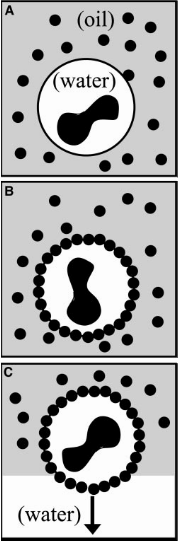
A single cell moves by detaching and reattaching from the substrate, or the surface it is on, as the cell  expands and contracts. This movement exerts forces on the substrate. (These forces can actually be measured directly –  this is the topic of a previous softbites post.) When many cells move together in a “cell sheet”,  their motion becomes more complicated. Not only do cells push and pull on the substrate, but they also push and pull on the cells that surround them.  In today’s study, Xavier Trepat and colleagues show that there is a “tug-of-war” between cells that causes them to migrate.

Previously, it was thought that only the cells at the very front of the mass of migrating cell, or the leading edge of the cell sheet, exert forces on the substrate. According to this picture, most of the cells get passively pulled along by the leading edge, and neither push nor pull on the substrate. By measuring the forces the cells exert on the substrate, Trepat and his colleagues discovered that, in fact, all of the cells are involved in pushing the cell sheet forward.

The researchers measured the forces in a moving sheet of cells, taken from canine kidneys, growing on a gel substrate using a technique called traction force microscopy. The first step of this technique is to track the displacements of different points within the substrate as the cells move. Then, the mechanical properties of the gel are used to calculate the forces on the substrate generated by this motion. The researchers mapped the value of these forces using different colors, with red and blue representing very strong forces and black representing zero force. They first looked at what happened at the leading edge of the cell sheet, as in Figure 1.


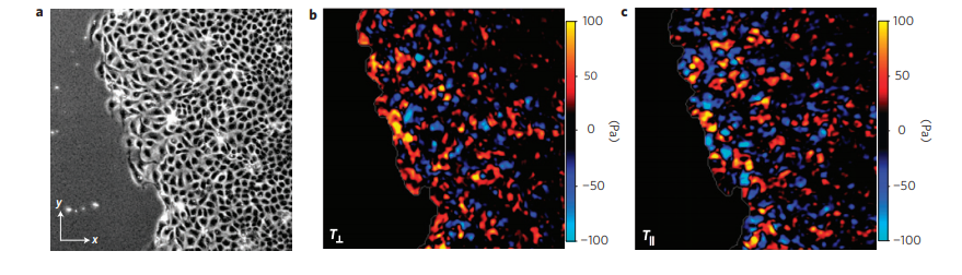
Figure 1. a. Image of the cell sheet, in which individual cells are outlined in white. The field of view is 700 microns by 700 microns. b. The forces that the cells exert perpendicular to the leading edge of the cell sheet. c. The forces that the cells exert parallel to the edge of the cell sheet. Bright red and blue colors indicate strong forces (up to 100 Pa of stress), while black color indicates low forces. (Images adapted from the original article.) The cell sheet’s expansion was recorded in a video as well.

The researchers separated the normal forces (Figure 1b) — those exerted by the cells perpendicular to the leading edge of the cell sheet, or in the direction of the cells’ motion — from the forces exerted parallel to the leading edge of the cell sheet (Figure 1c). The bright red and blue colors in Figure 1 show that cells well inside the cell sheet exert forces on the substrate. From this, they hypothesized that instead of having “follower” and “leader” cells, all the cells contribute into pushing and pulling the cell sheet as they move.

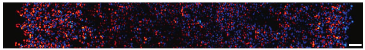
The researchers then looked at larger areas of the cell sheet, such as that  shown in Figure 2. The bright colors near the edges correspond to strong forces,  while the black spots show that the forces in the center of the cell sheet are weaker. This suggests that the cell sheet “tugs” both to the right and the left as it expands. As the cells exert forces on the substrate, they exert forces on each other. The cells pulling to the right and the left are similar to two teams pulling a rope in a game of tug of war. The sheet of cells is like a rope that grows in the direction of the tugging of the cells.

Figure 2. Forces exerted by a larger piece of the cell sheet. Bright red indicates strong positive forces and blue indicates strong negative forces, while black indicates low forces. The scale bar on the bottom right is 200 micrometers.  (Image adapted from the original article.)

Next, the researchers wanted to understand how being tugged on by its neighbors affects the motion of individual cells: does the tug of war consistently pull a cell in a particular direction? Or is the cell equally likely to be pulled in any direction?  To answer this question, Trepat and colleagues measured the average force exerted on a cell by its neighbors, as a function of the distance of that cell from the edge of the sheet. If each cell was moving independently, the average normal force inside the sheet would be zero – on average, no cell would be pushing or pulling any other cell to a specific direction. Instead, as shown in Figure 3, the average force was not zero, and was actually higher for distances farther from the sheet’s leading edge. In other words, the cell sheet is expanding from the inside more than it’s being pulled from the edge.


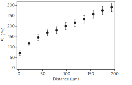
Figure 3. The average normal force exerted on a cell by its neighbors, \sigma_{xx}, is higher farther from the leading edge of the cell sheet. (Figure adapted from the original article.)

Each individual cell crawling on a substrate has little effect on its surroundings, but many cells acting together can exert forces on each other to guide the collective in a particular direction. As cells replicate, such as in a healing wound, this guiding helps the cells expand in directions where there is space to be filled. This study by Trepat and colleagues reveals for the first time the tug-of-war that allows the tissues in our bodies to grow and heal.

The Origin of Random Forces Inside Cells

Original paper: Probing the Stochastic, Motor-Driven Properties of the Cytoplasm Using Force Spectrum Microscopy 


Place yourself in a bumper car at a carnival waiting to bump into your friends. Soon enough you hear the small engine of your bumper car start and you begin to move around, bumping into anyone in your way. While the motion of your car is mostly controlled by the steering wheel, random events—like fluctuations in the motor power, your car hitting small bumps on the floor, and other cars hitting you—can affect the motion as well. What if I told you that a cell and its parts function in a similar way? Just as your car is powered by electricity, molecular motors—bio-molecules that can convert chemical energy into mechanical work—power the movement of living organisms by generating forces. In order to produce these forces, molecular motors depend on an organic molecule called ATP [Footnote: Adenosine TriPhosphate]. And just like the fluctuations in the motor power of the bumper car, random fluctuations can also be produced by the molecular motors.

The motion caused by molecular motors is necessary for the functionality of the cell—for example, division and contraction. However, it’s not this directed motion that’s studied in today’s paper, but rather the random fluctuations that accompany it.  But how can we extract useful data from random movements like those in the cytoplasm? One way is to measure the mean squared displacement (MSD) of a particle in the fluid. The MSD is a measure of how far a particle moves from its starting point over time. Going back to the example of the bumper car, you could find your MSD by tracing your path and seeing how far you have moved from your starting point over time1.


Screen Shot 2018-09-11 at 9.24.32 PM.png
Figure 1: Trajectories of particles inside a cell show Brownian-like motion.

To investigate the motion of particles in the cytoplasm, Guo and colleagues injected tiny particles into the cells and tracked their motion using confocal microscopy—a technique that allows for the precise tracking of the 3D position of micro-particles. After tracking the particles over time, Guo calculated the MSDs of the particles2.

Guo and colleagues observed that at short timescales, t ? 0.1s, the MSDs were nearly time independent, meaning that they did not change over time (see Figure 2A). This type of motion is typically observed in elastic solids, where particles can never move very far from their starting points. At longer timescales measured, 10s ?t ? 0.1s, the MSDs grew linearly with time. This type of motion is called Brownian motion and is usually observed in particles moving in viscous fluids under the influence of thermal forces. This association between linear MSDs and Brownian motion is strong enough that researchers have sometimes assumed that that particles inside cells move primarily due to thermal forces.  However, as discussed earlier, molecular motors generate forces inside the cytoplasm. Is it possible that random forces from molecular motors affect the motion of the particles?

In order to answer this question, Guo and his colleagues reduced the amount of ATP in cultured cells, thus reducing the activity of the molecular motors. They observed that the MSDs of particles inside ATP-depleted cell didn’t exhibit the linear MSDs seen in the untreated cells (see Figure 2B). This observation means that forces causing Brownian motion in the cytoplasm were ATP-dependent and therefore not generated by random thermal motion alone.


Screen Shot 2018-09-11 at 9.26.01 PM.png
Figure 2: MSDs of Microinjected Particles
(A) The average MSDs of different sized particles were plotted against time. On the plot, the dashed line corresponds to Brownian motion. (B) The MSDs normalized by particle diameter 3 in untreated (normal, ATP-containing) and ATP-depleted cells. The particles in ATP depleted cells move much less, and do not exhibit Brownian motion.


In short, Guo and colleagues showed that molecular motors impact the random motion inside the cytoplasm of a cell. The team proved this by measuring the MSD of particles inside cultured cells. They then depleted ATP in the cells to observe any changes in the MSDs of the particles inside. They found that movement inside the cytoplasm was largely affected by random molecular forces produced by molecular motors and not solely due to thermal forces.  However, this discovery raised more questions. For example: why do these molecular motors, which exert directed forces, exhibit random movements? We’ll answer this question in a follow-up post by considering the elastic network that couples molecular motors.


1. Note that a post by Christine Middleton has gone over a slightly different application of the MSD here: https://softbites.org/2018/04/25/the-matter-of-maternal-mucus-permeability-and-preterm-birth.?

2. Mean Squared Displacement: <?r2(?)> ; < ?r(?) > = r(t+?)-r(t) ?

3. The purpose of normalizing the data is to more easily compare the data between different particle sizes. ?

Brick-by-brick to Build Tiny Capsules

Original paper: Colloidosomes: Selectively Permeable Capsules Composed of Colloidal Particles


Disclosure: The first author of the article discussed in this post, Anthony Dinsmore, is now my Ph.D. advisor. He did his postdoc at Harvard University a while ago, and consequently, I was never involved in this work.

In past two decades, several approaches have been developed and optimized to encapsulate a wide variety of materials, from food to cosmetics and the more demanding realm of therapeutic reagents. Inspired by biological cells, the first attempts were to use either natural or synthetic lipid molecules to form encapsulation vessels, the so-called liposomes. Then, with the increasing awareness of controlled release of cargo, especially for therapeutic purposes, advanced materials such as polymers were developed to form carrying vessels. There has been an enormous progress in encapsulation technologies, however, these methods can be limited in their applicability regarding encapsulation efficacy, permeability, mechanical strength, and for biological applications, compatibility. In this article, Anthony Dinsmore and his colleagues introduce a new platform and structure to encapsulate almost all types of materials with finely controlled and tuned properties.

Colloidosomes

An emulsion is produced typically by application of a shear force to a mixture of two or more immiscible liquids like the classical water-oil mixture. The resulting solution is a dispersion of droplets of one liquid in the other continuous liquid. In such case, an interface between the fluids exists that would impose an energy penalty on the system. Therefore, the system will always attempt to minimize it, in essence by reducing the area of the interface that is to merge the similar liquid droplets. Amphiphilic molecules are known to segregate in such interface to further reduce the energy and to inhibit the merging of droplets.  This segregation is not limited solely to molecules though. Solid particles tend to jam in the interface for the same reason to stabilize the emulsions. Inspired by the idea of particle-stabilized emulsions, which are known as Pickering emulsions, Dinsmore, and his colleagues have developed capsules made of solid particles. They adopt the name “Colloidosomes” by analogy to liposomes and demonstrate how the arrangement of these particles can be manipulated and controlled to achieve a versatile encapsulation platform.

Fabricating the Capsules

Colloidosomes are prepared first by making the emulsion in which the continuous phase contains the particles. For instance, in water-in-oil emulsions (“w/o”), water droplets become the core of the colloidosomes and particles are dispersed in the oil phase. Gentle agitation of such system results in particles being trapped in the water-oil interface (see Fig.1). The authors summarize the capsule formation in three main steps:


Screen Shot 2017-10-17 at 01.01.10
Fig 1. The colloidosome formation process is illustrated schematically in three steps. (A) a water/oil emulsion first is created through gentle agitation of the mixture for several seconds. (B) Particles are adsorbed to the w/o interface to minimize the total surface energy. Through sintering, van der Waals forces, and or addition of polycations ultimately the particles are locked in the interface. (C)In the end, the particle-stabilized droplet is transferred to water via centrifugation.

(a)  Trapping and stabilization. When the water-oil interface energy surpasses the difference between particle-oil and particle-water interface energy, particles are absorbed to the water-oil interface and become trapped due to the presence of a strong attractive well. This differs substantially from the case where particles were adsorbed to the interface via electrostatics, which requires the droplets to be oppositely charged to attract the particles. The packing of the particles at the interface is adjusted by controlling their interactions. Typically, the electrostatic interaction between particles, due to their surface chemistry, is utilized to stabilize the packing of the particle. For instance, in this study particles are coated with a stabilizing layer which in contact with water turns into a negatively charged layer.


(b)  Locking particles. To form an elastic and mechanically robust shell, the particles must be locked in the interface. This results in an intact capsule that can withstand mechanical forces. One way to obtain such elastic shell is to sinter the particles in place. Sintering is a thermally activated process in which the surface of particles melts and connects them to each other. Upon this local melting, the interstices among particles begin to shrink. With longer sintering times, it is possible to completely block the interstices, which results in very tough capsules with extremely high rupture points.  In this study, particles with 5 minutes of sintering yielded a 150 nm interstices size, and with 20 minutes, almost all the holes were blocked. By using particles with different melting temperatures, the sintering temperature can be adjusted over a wide range; this might be advantageous for encapsulants incompatible with elevated temperatures. Other ways of locking particles are electrostatic particle packing and packing via van der Waals forces. In the former case, for instance, a polyelectrolyte of opposite charge can be used to interact with several particles to lock them in place. In the latter case, for the van der Waals force to be effective, the steric repulsions and barrier must be destroyed so the surface molecules can get close enough for the London forces [1] to be strong.


After the Colloidosomes are formed, through gentle centrifuging, the fluid interface can be removed by exchanging the external fluid with one that is miscible with the liquid inside the colloidosome. In this step, having a robust shell to withstand shear forces crossing the water-oil interface is very important. This process ensures that the pores in the elastic shell control the permeability by allowing exchange by diffusion across the colloidosome shell. Now, with these steps and knowing parameters such as surface chemistry and locking condition, a promising system with characteristic permeability or cargo release strategies can be designed.


Tuning Capsule Properties; Permeation and Release

The most important feature of a colloidosome, as a promising encapsulant, is the versatility of permeation of the shell and or the release mechanisms. Sustained release can be obtained via passive diffusion of cargo via interstices that can be tuned via particle size and the locking procedure. With the mechanical properties of capsules optimized, shear forces can be used as an alternative release mechanism. For instance, minimally sintered polystyrene particles of 60 microns in diameter have shown to rupture in stresses that can be tuned by sintering time over a factor of 10. What makes the colloidosomes even more interesting is that one can choose different particles, with different chemistry, to have an auxiliary response, such as swelling, and dissolving of particular particles in response to the medium. It is also conceivable if one coats the colloidosome with the second layer of particles or polymers to improve or sophisticate the colloidosomes response. The latter can also mitigate the effects of any defect in the colloidosome lattice.

        With this unique platform, Dinsmore and colleagues stepped into the new realm of encapsulating materials of all kind. From therapeutic cargos to bioreactors, the chemical flexibility and even the ease of post-modification would expand the cargo type beyond molecules. For example, the authors show that living cells can be encapsulated in colloidosomes. Well, you may wonder, WHY? Imagine a protective shell around cells that keep them out of the reach of hostile microorganisms without compromising the cell’s vital activities such as nutrient trafficking and cell-to-cell crosstalk. 


[1]  London forces arise when the close proximity of two molecules polarizes both molecules. The resultant dipole work as a magnet to glue molecules together. Therefore, London forces are universal forces (and part of van der Waals forces), which takes effect when atoms or molecules are very close to each other.