
Swat it or Print with it? The story of a mosquito’s second life!

Original paper: 3D necroprinting: Leveraging biotic material as the nozzle for 3D printing

Author: Manikuntala Mukhopadhyay
Editor: Jack Llewellyn


If there is one insect that rarely receives any appreciation, it is probably the mosquito! Mosquitoes are infamous for ruining summer evenings, causing itchy bites and spreading some of the world’s deadliest diseases. Most of us spend our lives trying to avoid them. However, a team of researchers from Canada’s McGill University recently found an unexpected use for one of nature’s most unpopular insects: turning mosquitoes into components of a 3D printer.

Modern 3D printers often work by squeezing material through a nozzle, rather like icing being piped onto a cake. The smaller the nozzle, the finer the structures that can be printed. However, manufacturing extremely small dispensing nozzles is neither simple nor sustainable. High-resolution dispensing tips are typically made from metals, plastics, or glass and require specialised fabrication techniques that can be expensive, technically challenging and energy intensive.This led the researchers to ask a rather unconventional question: what if nature had already engineered an alternative?

Rather than building a new nozzle from scratch, the researchers decided to borrow one from nature, specifically, from a mosquito. To do so, Changhong Cao and his team introduced a technique called 3D necroprinting, in which parts of deceased mosquitoes were repurposed as engineering components. Although we usually think of mosquitoes as little more than irritating bloodsuckers, their feeding apparatus is a remarkable feat of natural engineering. The mosquito feeds through a needle-like structure called proboscis, a highly specialised microfluidic device refined through millions of years of evolution. To successfully feed on blood, the proboscis must satisfy several demanding requirements. It needs to be stiff enough to penetrate skin, thin enough to minimise damage, and hollow enough to transport fluid efficiently. Most importantly, its internal diameter is only about 20–25 micrometres, roughly one quarter the width of a human hair. In other words, the mosquito already possesses the kind of microscopic fluid-delivery channel that engineers spend considerable time, effort and resources trying to fabricate.
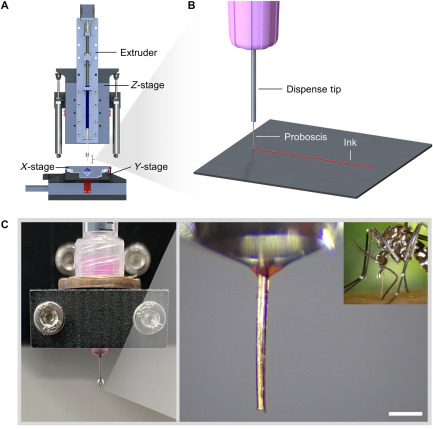
Figure 1: (A) Schematic of the custom-built 3D printer used in the study. (B) Illustration of the printing concept, where a mosquito’s proboscis acts as an ultra-fine dispensing nozzle. (C) Experimental setup showing a mosquito proboscis attached to a conventional printer tip. Scale bar: 50 micrometres

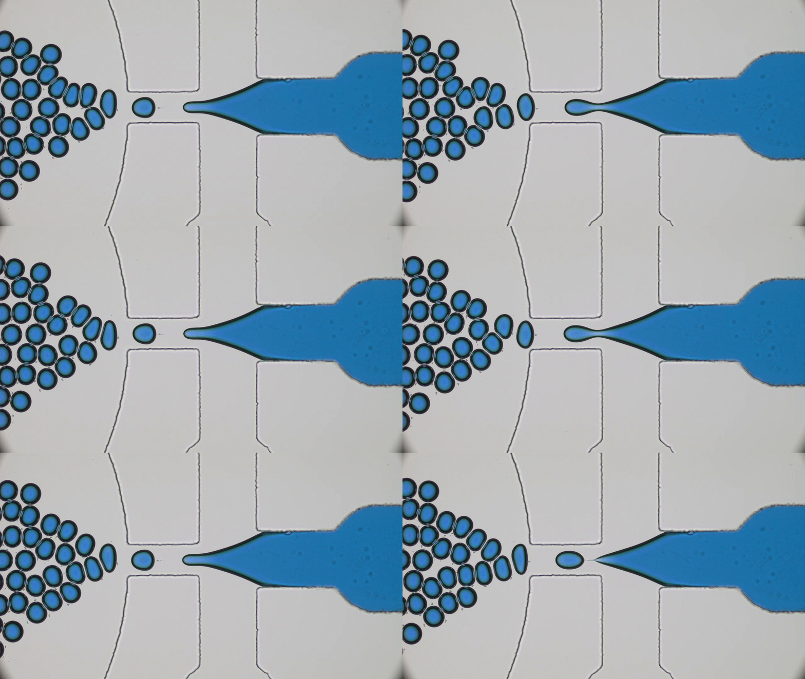
Next, to turn a mosquito into a printer nozzle, the researchers carefully extracted the feeding tube from female mosquitoes and attached it to a 3D printer. The concept was very simple. Figure 1 shows how instead of ending with a conventional metal nozzle, the printer terminated in a mosquito proboscis. Ink was pushed through the biological channel (Figure 2) and deposited onto a surface in precisely controlled patterns.
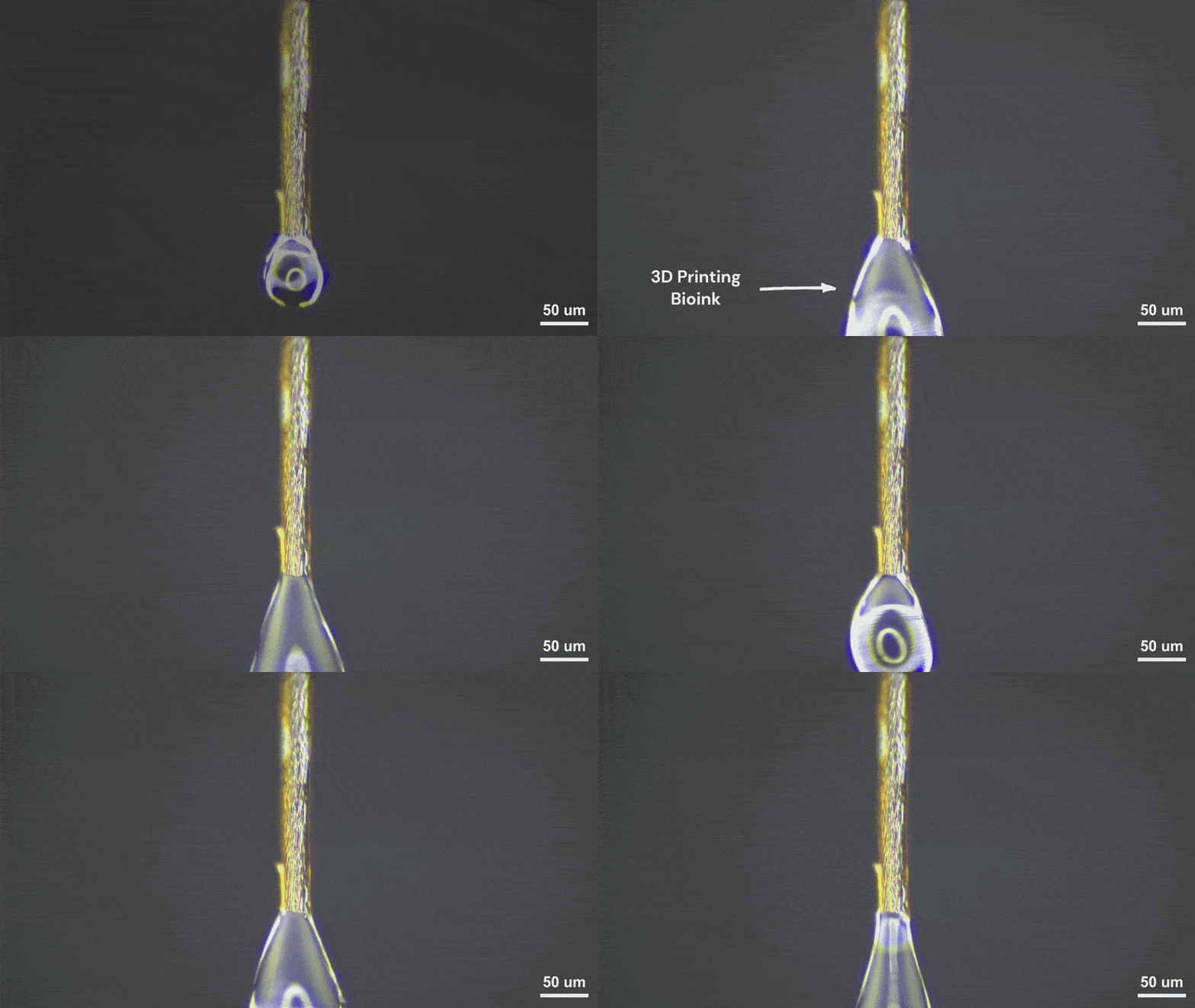
Figure 2: Printing with a mosquito proboscis nozzle

Although mosquito mouthparts appear delicate enough to break at the slightest touch, they are mechanically tougher than one might expect. To test their strength, the researchers sealed the tip of the proboscis and gradually increased the internal pressure, much like inflating a tiny balloon until it bursts. Surprisingly, the proboscis withstood pressures of approximately 60 kilopascals before rupturing. This was sufficient for many common printing inks used in bioprinting applications.

Once the mechanical limits had been established, the researchers began printing. The results were impressive! Using the mosquito nozzle, the team produced printed lines as narrow as 18–28 micrometres, substantially finer than those typically produced using common commercial dispensing tips.  To demonstrate the capabilities of the system, they also fabricated a range of intricate structures including microscopic honeycombs and maple leaves (Figure 3). After all, what better way for a Canadian team to demonstrate a new printing technique than by printing a tiny maple leaf?
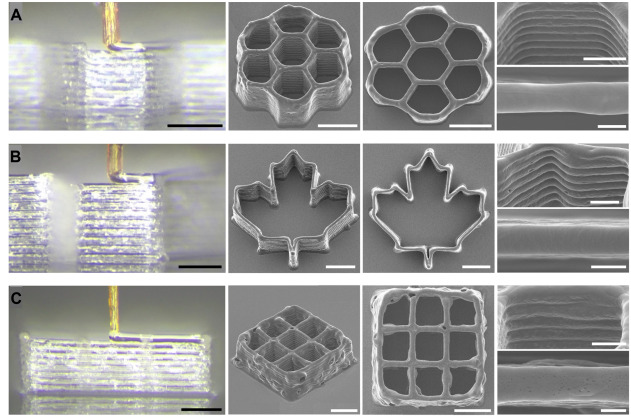
Figure 3: The mosquito proboscis was used to print a variety of microscopic 3D structures, including (A) a honeycomb pattern, (B) a maple leaf design, and (C) a cell-laden scaffold. Scale bars from left to right, 100, 200, 200, and 200 micrometres; top, 100 micrometres; and bottom, 20 micrometres.

Perhaps the most exciting aspect of the work lies in its potential biomedical applications. The research team loaded their printing inks with living cells to show that the mosquito nozzle could handle delicate biological materials without causing excessive damage. This opens intriguing possibilities for future applications in tissue engineering, drug delivery and bioprinting.  In other words, the same biological structure that evolved to extract blood from living organisms might one day help fabricate tissues for regenerative medicine.

At first glance, 3D necroprinting may seem like a quirky scientific curiosity. Yet beneath the unusual headline lies a powerful idea. Nature has spent millions of years refining structures that are remarkably specialised. The mosquito proboscis is one such example! The next time a mosquito lands on your arm, you will probably still swat it. But before you do, it might be worth remembering that hidden inside that tiny insect is a remarkably sophisticated piece of engineering. This work reminds us that nature is not merely a source of inspiration, but also a vast library of ready-made solutions waiting to be discovered. Sometimes, the next breakthrough in engineering may already be buzzing around us!

Disclosure: The author declares no competing interest.
 

Untangling the mystery of shape-morphing worm swarms

Original paper: Ultrafast reversible self-assembly of living tangled matter

Author: Sri Ganesh Subramanian
Editors: Jack Llewellyn, Manikuntala Mukhopadhyay


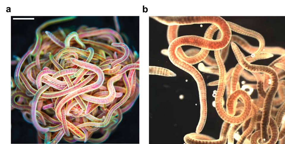
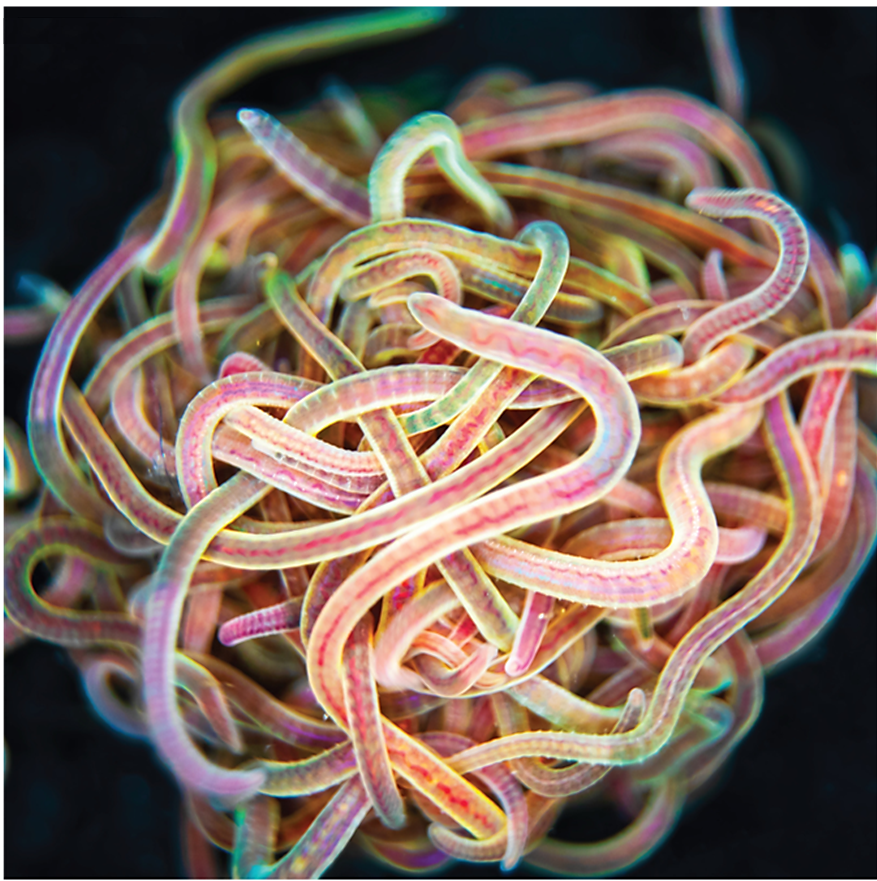
Many of us would have tied several knots very easily, but we all remember the frustration when trying to untangle the cords of a headphone – an experience that often tests our patience. Now imagine a creature that not only creates a tangled ball out of itself and its companions in minutes but can also untangle the entire knot in milliseconds if it senses danger. Meet the California blackworm (Lumbriculus variegatus), a small yet remarkable organism that is teaching researchers innovative lessons about managing tangles (see Figure 1).

Figure 1: (a) A compact, ball-like structure formed by California blackworms (Lumbriculus variegatus), demonstrating their natural tangling behavior. (b) The same blackworms rapidly untangle and disperse in response to a perceived threat. Scale bar: 3 mm.

At first glance, studying the movement of worms might seem unusual or even unpleasant. However, researchers in the Bhamla Lab at Georgia Institute of Technology and Massachusetts Institute of Technology, USA, were captivated by the rapid untangling behavior of these worms. The scientists noticed that these worms naturally tangle together for survival. By assembling themselves in the shape of a compact ball, they reduce water loss, regulate temperature, and protect themselves from environmental challenges. Their most astonishing trait, however, is their ability to instantly untangle (within a few milliseconds) and scatter in random directions when they feel threatened. Scientists are studying these worms to uncover how their movements enable them to form and break apart tangles so efficiently, and how these principles can be applied to develop innovative materials and technologies.

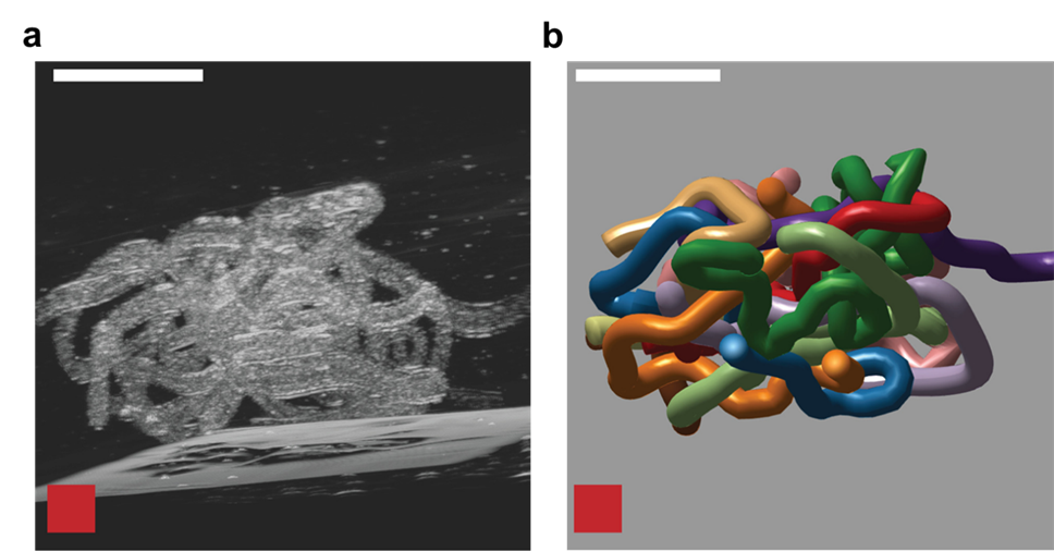
To understand how blackworms form and break tangles, scientists used ultrasound imaging to look inside these clusters. Because blackworms untangle almost instantly when disturbed, the researchers needed a method to slow their movements without harming them. The scientists carefully submerged the worms in a glycerol bath (a non-toxic viscous liquid commonly used in products like chewing gums and marshmallows) at a controlled temperature, to reduce their speed (Figure 2a). After allowing the worms to settle in the glycerol bath, the researchers carefully directed ultrasound waves from multiple directions to track the position of these worms as they moved over one another. This allowed the researchers to create a 3D image of the worms in their tangled state (Figure 2b). What they found was surprising: the tangles aren’t random. The worms were tightly packed, and most of them were in contact with one another, forming an intricate and highly organized structure.

Figure 2: (a) Ultrasound image of a tangled ball consisting of 12 California blackworms immersed in a glycerol bath. (b) Three-dimensional reconstruction of the ultrasound data, showing the positions of individual worms and their interactions within the tangle. Scale bar: 5 mm

By analyzing how the worms moved and interacted, researchers identified specific patterns that dictate how these tangles form and function. They discovered that each worm’s position and movement contribute to the overall structure. Additionally, the size of the tangle could range from a handful of worms to several hundred, depending on environmental conditions and the size of the group. The researchers also discovered that the movements of the worms are critical to their tangling abilities. To tangle, the worms moved slowly and created loops that naturally intertwine. Over time, these loops formed a dense and stable structure. Untangling, however, is a completely different process. When blackworms sense danger, they generate rapid, wave-like motions through their bodies known as helical waves. These waves act like a zipper being undone, loosening the knots and allowing the worms to quickly break free, within a few milliseconds.

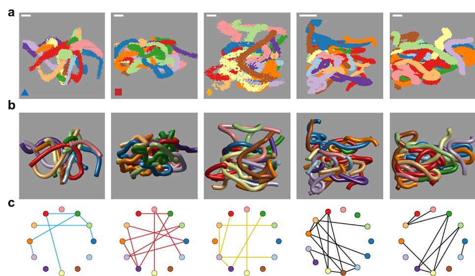
To better understand these motions, researchers created mathematical models to understand how the movement of the worms lead to tangling and untangling. The models showed that factors such as speed, direction, and frequency of the worms’ movements are precisely tuned to achieve their goals with remarkable efficiency. One of the most exciting outcomes of this research is the creation of a “tangle map” (see Figure 3). This map identifies the factors that influence whether worms tangle or untangle. These factors include movement speed, the angles at which the worms twist, and how often they change direction. By manipulating these variables, the worms can control whether they form a strong tangle or quickly break free and disperse.

Figure 3: (a) Data generated from ultrasound images show how groups of California blackworms tightly pack together. (b) Scientists used a method called the “diffusion maps algorithm” to trace smooth curves through the worm positions in the ultrasound images, making it easier to track each worm. (c) Tangle graphs were created from these ultrasound datasets to show how the worms are connected and tangled. Scale bars represent 2 mm.

This map also helps scientists understand how to replicate these behaviors in synthetic materials. For instance, by mimicking the worms’ twisting and untwisting motions, engineers could design materials that assemble and disassemble in response to environmental factors, such as temperature changes or mechanical stress. For example, in disaster response scenarios, robots inspired by blackworms could navigate through rubble by tangling and untangling themselves to squeeze into tight spaces. Similarly, materials that adapt to different conditions could improve everything from clothing to architecture. Blackworms remind us that sometimes the solutions to our most complex challenges are hidden in the simplest of creatures.

Disclosure: The author declares no competing interest.