“Spleen-on-a-chip” gives an inside view of sickle cell disease

Original paper: Microfluidic study of retention and elimination of abnormal red blood cells by human spleen with implications for sickle cell disease 

Content review: Arthur Michaut
Style review: Arthur Michaut


Though they may not realise it, anyone who’s taken the subway at rush hour knows how a red blood cell feels passing through the human spleen. Almost home now, just need to get through the gates; but wait, someone’s ticket isn’t working, the crowd is starting to push, the gates are getting jammed… Maybe you should have called a cab.

Figure 1. Left: in vivo filtration of red blood cells through the slits of the spleen. Right: in vitro filtration of red blood cells through silicone slits.

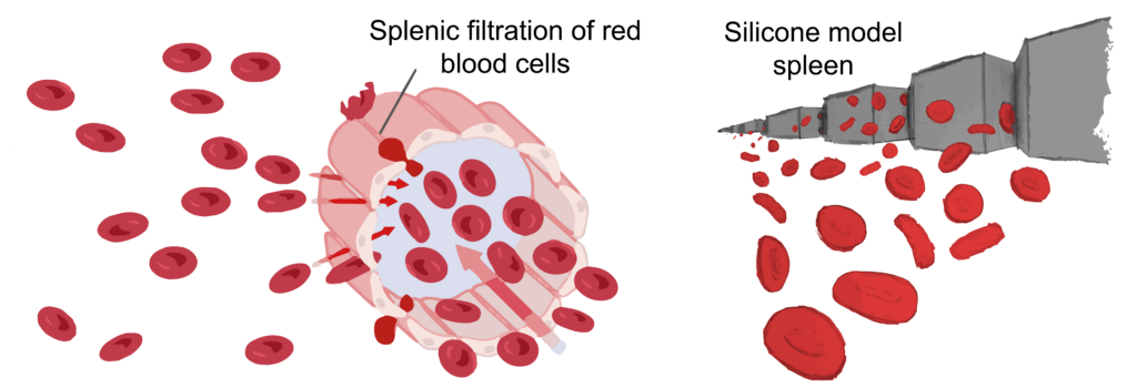
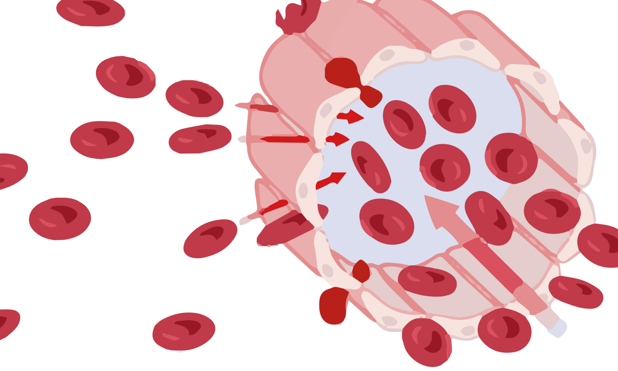
Much like turnstiles validating your ticket, the human spleen contains narrow slits that red blood cells must squeeze through to prove they are fit to navigate tight blood vessels while carrying oxygen around the body. The spleen is tasked with ensuring our blood contains the right amount of red blood cells and removing any cells that aren’t in tip-top shape. Shape, however, can be a problem the spleen can’t handle.

People with sickle cell disease produce “sickle” shaped red blood cells due to an inherited genetic mutation. Sickle cell disease patients can experience episodes of acute splenic sequestration, or “spleen crisis” where these misshapen cells clog the slits of the spleen, depriving it of oxygen and leading to swelling that can become life-threatening. Because this process is difficult to observe and monitor in the body, there is little understanding of how and why a spleen crisis might occur. When a spleen crisis occurs, an urgent blood transfusion is needed to treat the problem, however, in some cases, it becomes necessary to surgically remove the spleen.

A group of scientists spanning the USA, France, and Singapore have teamed up to understand how sickle cell disease can become life-threatening. Their device, a silicone model spleen, could be used to predict complications of the disease as well as develop new treatments.

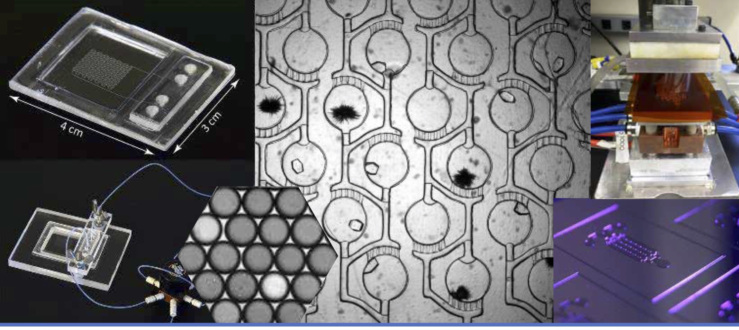
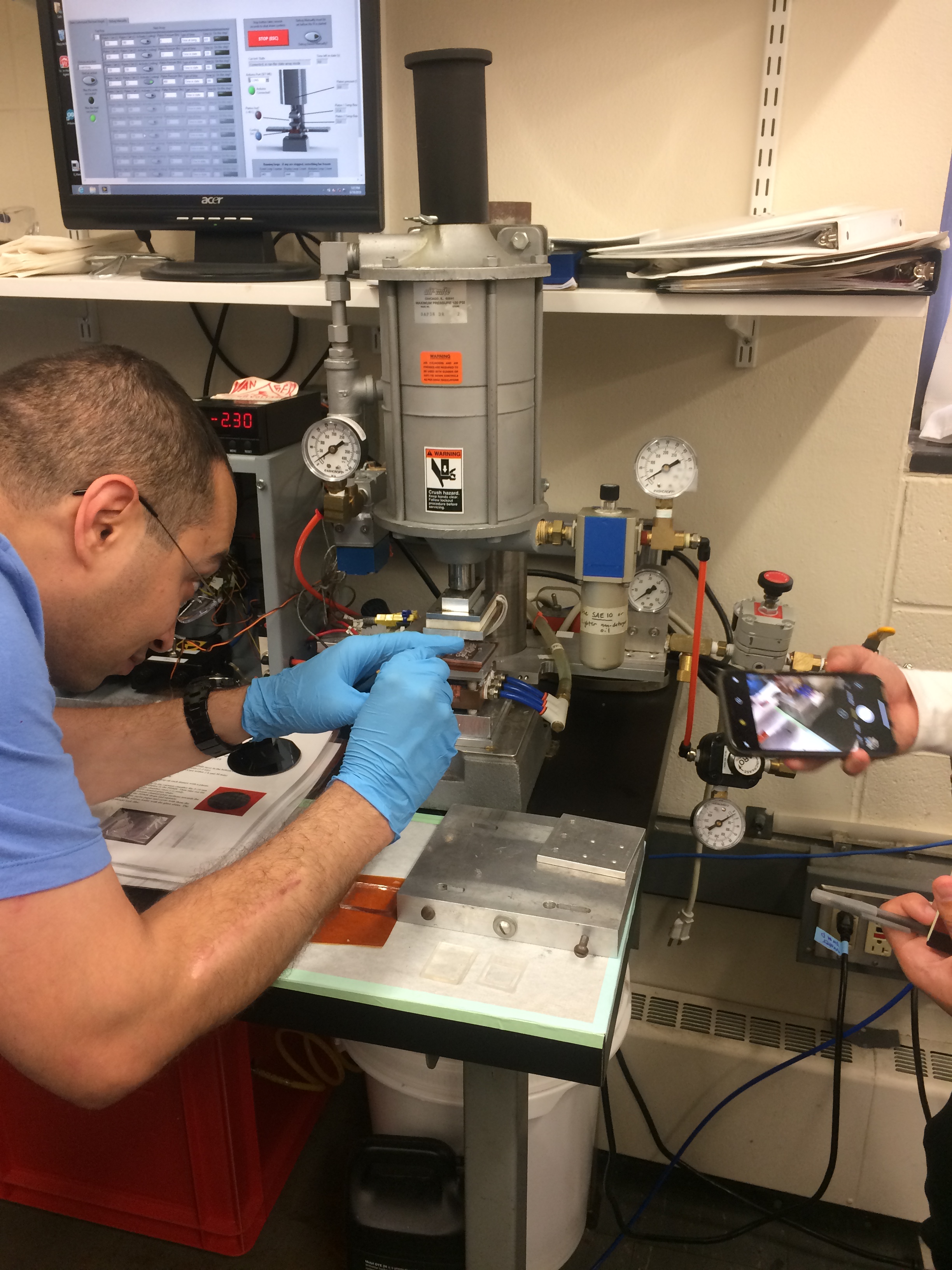
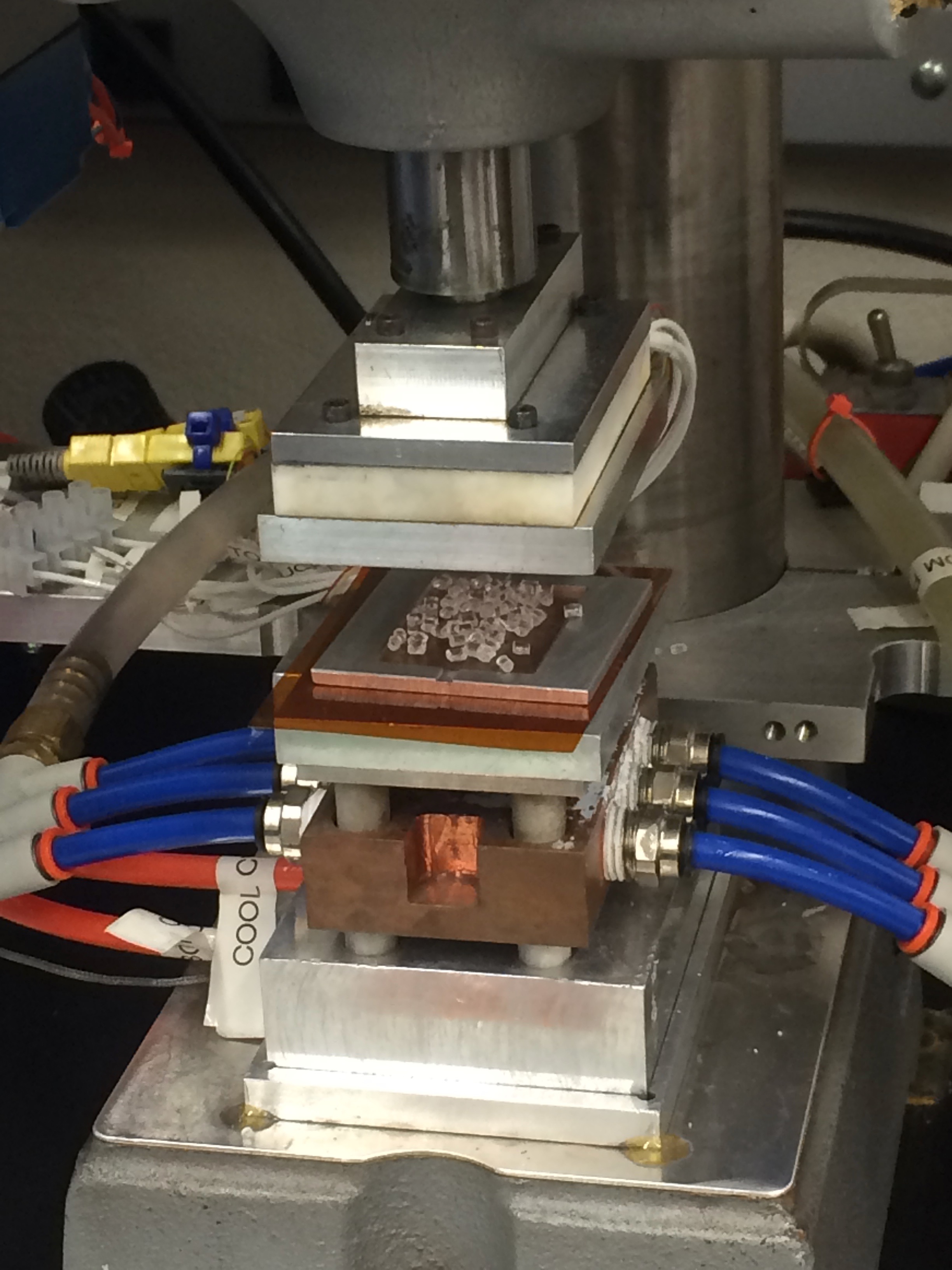
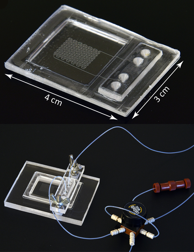
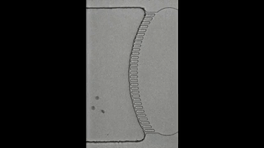
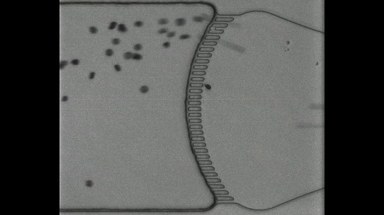
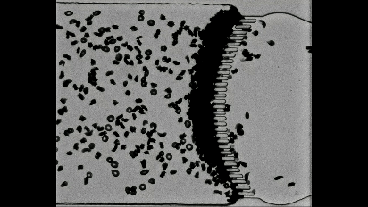
Using a soft material classically used in microfabrication, named PDMS, to cast a mold with slits similar to those in the human spleen (Figures 1 & 2i), researchers were able to directly observe the processes that lead to spleen crises. The group analysed flows of red blood cells from both healthy and sickle cell disease patients through the silicone spleen, simulating splenic blood flow rate and oxygen levels, and measured retention of cells in the slits of the device.

The flow of red blood cells from healthy and sickle cell disease patients through the silicone spleen.
Figure 2. (i) The biomimetic “spleen-on-a-chip”, scale bar 10 µm (with higher magnifications outlined in red, right). The majority of red blood cells from healthy patients (ii) pass through the device, however, cells from sickle cell disease patients (iii) are more frequently retained at the slits of the device, and when deprived of oxygen (iv) these cells block the device entirely.

After one minute of blood flow, red blood cells from sickle cell disease patients blocked more than double the number of slits as healthy red blood cells (Figures 3i & 3ii), demonstrating how the spleen can become blocked and swollen in sickle cell disease patients. When the silicone spleen was deprived of oxygen, as occurs during a spleen crisis, red blood cells from sickle cell disease patients became stiffer, more viscous, and more frequently sickled (Figure 3iii). Under this condition, sickled red blood cells completely blocked all of the slits. Upon reintroducing oxygen into the system, many cells return to their normal shape and blockages begin to open up again within seconds (Figures 3iv & 4).

The effects of de- and re-oxygenation on red blood cells from healthy and sickle cell disease patients and the unblocking of the device following reoxygenation.
Figure 4. Left: red blood cells from healthy (AA) and sickle cell disease (SS) patients under oxygenation and deoxygenation conditions (scale bar 10 µm). Right: the percentage of open slits in the device quickly increases when oxygen is reintroduced into the blocked system.

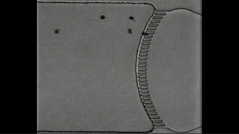
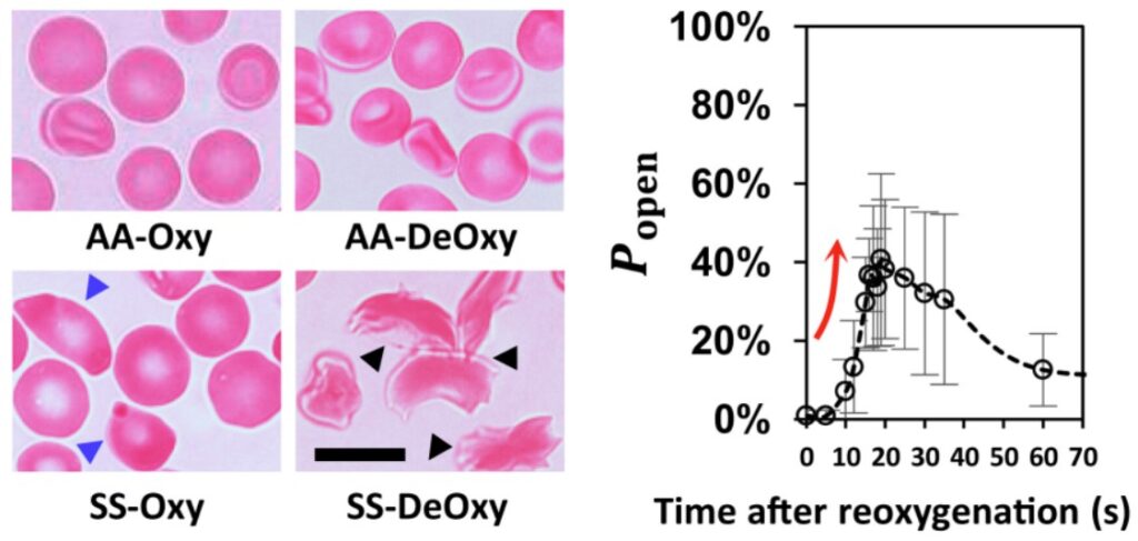
The results uncover a viscous cycle leading to spleen crises in sickle cell disease patients. Sickled cells cause blockages in the spleen, which deprives the spleen of oxygen, which in turn causes more cells to become sickled. Additionally, it was shown that the addition of oxygen caused cells to “unsickle” and rapidly cleared blockages in the slits, revealing why immediate blood transfusion can alleviate spleen crises. The researchers speculate that a sudden burst of oxygen-rich red blood cells via transfusion has the same effect as when they reintroduced oxygen to their blocked system.

Researchers believe the spleen-on-a-chip could become a new tool to allow sickle cell disease patients to monitor their condition and allow for early diagnosis of spleen crises, as well as providing a testing ground for new treatments against the disease. Safe travels red blood cells!

Disclosure:  The author declares no competing interest.

2019 MRSEC Brandeis Microfluidics summer course

If you just landed on Softbites for the first time, you probably have not had the chance to read our previous posts about microfluidics (like this one, or that one, and more). If this field of science is foreign to you, all you need to know is that it studies how fluids flow at really small scales (typically tens to hundreds of micrometers). For instance, you can quickly generate tiny droplets of a solution, turning each droplet into an individual “reactor”. Or you can create  microenvironments with precisely controlled chemical concentrations to grow cells in different conditions.

In addition to being a thriving field of research, I think microfluidics is simply beautiful! I have spent hours looking at the Softbites website’s banner, a movie of droplets that was shot by the Lutetium project. You can imagine my excitement when I registered to the annual MRSEC microfluidics summer course 2019 at Brandeis University. This summer course was run by four talented grad students from the Fraden lab and the Rogers lab: Ali Aghvami, Alex Hensley, Marilena Moustaka and Zahra Zarei. These labs are part of the MRSEC program at Brandeis, an important place in the New England soft matter community. Therefore, I think it was the perfect place to get started with microfluidics!

Figure 1. Me, trying to pour some resin on a silicon wafer (left). A drop maker setup (middle). A gradient maker chip, with a defect leading to a non-stable gradient (right).

Over five days, we learned the basics of one of the standard methods for making microfluidic channels, called soft lithography. The rationale is to make a mold using a UV-light sensitive resin. A 2D pattern can then be polymerized in the resin by shining UV-light through a mask. Whatever the UV light hits gets hard, while the rest of the resin stays soft. The soft resin is washed away leaving only the hard, UV-treated resin behind in the shape of the mask. The mold will finally be used to imprint the design in a soft transparent material called PDMS (a very nice video from the Lutetium project explains all this process). We experimented with this fabrication during three main sessions: 

  1. We drew our 2D design using a drawing software 
  2. We fabricated our mold in a clean room so no dust ruined our tiny features
  3. We cast the PDMS on the mold and sealed the device with a glass slide

We were taught each of these steps through a combination of lectures and hands-on sessions. You can see a droplet maker and a gradient making device that we made in Figure 1. 

In addition to learning how to make these routinely used PDMS-based devices, we were also introduced to another technique used in the Fraden lab. This technique, which was recently published (2017), uses a thermoplastic (a plastic that melts at moderate temperatures) as the main device material. This thermoplastic can be cast onto a PDMS mold by means of a thermopress (as shown in Figure 2). Unlike PDMS, thermoplastic is not permeable to water and organic solvent, and is stiffer. If the permeability of PDMS is a limitation for your microfluidics application, thermoplastic might be the way to go!

Figure 2. Ali Aghvami placing thermoplastic chips onto the PDMS mold (left). Close-up on the thermoplastic in the thermopress before being cast (middle). The final device (right), from Aghvami et al. 2017.

This week-long course introduced us to both classical microfluidics techniques that are routinely used in labs and some more advanced ones. More importantly, our instructors dedicated important time to discuss our personal projects with us. We even had a consulting session with Seth Fraden! I strongly encourage anyone to attend the next editions of this course. Each year, the dates and the call for applications are released in spring, so don’t miss out!

These were my first drops! I literally spent 45 minutes watching them!