The key to fighting cancer: be flexible

Original paper: Nanoparticle elasticity directs tumor uptake 


In my previous post on soft nanoparticles, you were introduced to polymer-based nanoparticles that could be used in biomedical applications, one of which is cancer therapy. These nanoparticles have a range of useful properties for cancer treatments, including their spherical shape and small size (~100 nm), both of which are similar to exosomes, small globules that are used in nature for transferring proteins between cells. Since cells naturally absorb exosomes, artificial particles with this size and shape should also be easy for cells to absorb, which means these particles could be used to deliver drugs into cells. While this idea sounds promising, it hasn’t worked out in practice —  when drug-loaded polymer-based nanoparticles were injected into a tumor, subsequent tests showed that less than 1% of the injected dose entered the cancer cells. Since these particles were the correct size and shape, why didn’t they get inside the target cells?

One possibility is that the elasticity (or stiffness) of nanoparticles is to blame: scientists have suspected that this mechanical property can affect the ability of nanoparticles to squeeze themselves through the cell’s membrane. Unfortunately, it is difficult to test this hypothesis directly, because modifying the elastic properties of a nanoparticle generally requires modifying its chemical properties as well. To solve this problem, Peng Guo and coworkers designed a special kind of nano-objects — spherical nanolipogels — with tunable elasticity. In this paper, they proved for the first time that breast cancer cells take up soft, squishy particles more easily than they take up hard ones.  

So what are nanolipogels? This type of nanoparticles is basically an altered version of a nanoliposome, a particle-like object that consists of a liquid water core surrounded by a layer of phospholipid molecules [1]. Guo and his colleagues created nanolipogels by filling the nanoliposomes’ liquid core with a polymer of tunable chemical structure. Nanolipogels have precise size (160 nm) and shape (spherical), and their elasticity can be made to vary without changing their other properties (see Figure 1).

image 1
Figure 1. Structures (top) and micrographs (bottom) of nanoliposomes and nanolipogels of increasing stiffness (higher values of Young’s modulus). (Image adapted from Guo’s paper.)
figure 2
Figure 2. Experimental setup of an Atomic Force Microscope. The height of a sample’s surface is scanned by a tip on a moving cantilever and the cantilever deflections are detected by a laser light to give the samples topographic profile. (Image from simple.wikipedia.org)

To measure the elasticity of the particles they had produced, Guo and coworkers used a technique called Atomic Force Microscopy (AFM). AFM is commonly used to visualize soft materials by imaging the height of their surface through the deflection of a cantilever (Figure 2). In this paper, the researchers used AFM for a different purpose: to calculate the Young’s modulus — a measure of stiffness — of the nanoparticles. They did this by compressing the particles between the cantilever tip and a solid surface, allowing the researchers to measure the force required to deform the particles by some known amount. The relationship between the applied force, the degree of deformation, and the Young’s modulus is given by the Hertz equation [2]. What you need to remember is that the greater the modulus, the stiffer the particle.

The researchers created four different nanolipogels of different elasticity with Young’s moduli ranging from 1.6 MPa (roughly the stiffness of cork) to 19 MPa (the stiffness of leather), and a nanoliposome without polymer in the core with a Young’s modulus at 0.045 MPa (roughly the stiffness of gummy bears). After verifying that all 5 particles could successfully encapsulate drug molecules, they tested how well tumor cells could uptake each particle. To do so, they used breast cancer cells in the lab (in vitro cellular uptake) and attached fluorescent dye to the particles to determine whether they were inside or outside of the cells. They found that the stiffest nanolipogels were 80% less effective compared to the softest nanoliposome samples; in other words, five times more of the softer particles got inside the cells. In vivo tumor uptake studies, using live mice, similarly showed that the nanoliposomes had up to 2.6 times higher cellular uptake than the stiffest nanolipogels.

Why do the soft nanoliposomes enter the cells more easily? To understand the conclusion of Guo and colleagues, we need to think about how nano-objects enter a cell. Figure 3 shows two possible ways of doing this: 1. fusion, where nano-objects break up and join the cell membrane, or 2. endocytosis, where the whole object enters the cell by bending the cell’s membrane and getting covered in a membrane outer layer. Fusion needs less energy compared to endocytosis, where cell membrane bending and surface tension increase the energy. The researchers hypothesized that nanoliposomes use both fusion and endocytosis, with a preference for fusion (Figure 3a), while nanolipogels can only enter the cell through endocytosis (Figure 3b). This hypothesis was verified by using chemical compounds that prevented endocytosis from taking place; in all experiments, the cellular uptake of nanoliposomes was as high as before, while much fewer nanolipogels were detected in the cells, since they couldn’t enter through endocytosis.

image 3
Figure 3. The possible pathways of (a) nanoliposomes and (b) nanolipogels entering a cell. (Image adapted by the Guo paper.)

This study showed that a nanoparticle’s mechanical property, in particular, its elasticity, affects how it enters cells, a finding that could potentially have a tremendous impact on cancer treatment and diagnosis. The use of nanoliposomes, which are a synthetic equivalent of nature’s drug delivery systems, may also be used in the future to further understand how cellular processes, such as fusion and endocytosis, take place.


Continue reading “The key to fighting cancer: be flexible”

3 Easy Steps to (almost) Curing Type 2 Diabetes

Original paper: Synthetic beta cells for fusion-mediated dynamic insulin secretion


Type 2 diabetes currently affects ~ 410 million people worldwide. It is a chronic condition caused by dysfunctioning beta cells in the pancreas. Beta cells normally secrete insulin in the pancreas to regulate blood glucose levels, and the loss of beta cell function can lead to hyperglycemia (i.e. high blood sugar), a condition with complications such as blindness and heart disease. The traditional invasive treatment involving direct insulin injection is a painstaking, never-ending process as it doesn’t properly regulate the dynamics of beta cells, just treats the symptoms. Modern treatments involve cell therapy in which functioning beta cells are transplanted from a healthy person, but this therapy faces serious challenges such as finding the right donor and suppressing the immune system after the transplantation. In this post, you will read how Chen and co-workers design an artificial version of beta cells that bypass the shortcomings of conventional cell therapy.

In our body, beta cells in the pancreas are responsible for monitoring and balancing our blood sugar level. When glucose levels are low (hypoglycemia, low sugar levels), these cells rapidly secret a polymeric (see note [1]) form of glucose. With high glucose levels (hyperglycemia) a hormone called insulin is secreted to bring down the concentration of sugar in the blood. Any disturbance to these cells, either through the body attacking itself (Type I diabetes, see note [2]) or genetic risk factors, can compromise the function of the beta cells, resulting in hypo- or hyperglycemia.

A4-fig1
Figure 1. The schematic of the vesicle-in-vesicle system. The giant micron-sized lipid vesicle (the OLV) encapsulates the machinery components for the insulin release, including the smaller vesicles that carry the insulin (ISVs). The image is taken from Chen and colleagues.

In this study, the researchers mimic the cell’s machinery in beta cells that sense sugar and signal a response inside the cell. This artificial system features two differently sized lipid vesicles (see Figure 1). The larger vesicle is about a micron in size (millionths of a meter) and is called the outer layer vesicles (or OLVs). It acts as the body of the artificial beta cell, encompassing the necessary machinery to regulate the insulin release. The second, smaller lipid vesicles, a thousand times smaller than the OLVs, are held inside the OLVs and are thus called inner layer small vesicles, or ISVs. These ISVs encapsulate the insulin hormone inside.

The system acts like a computer code with a conditional “IF” command to decide whether it needs to respond or stay inactive. IF the glucose levels outside of the OLVs are normal or below normal, no signal is induced. However, IF the glucose level increases beyond the signal-inducing concentration (which can be easily tuned by chemical modification, see below) then the signal is triggered, resulting in insulin release. The entire system consists of machinery to perform three distinct steps.

The first step is the glucose sensing step. Using a glucose transporter membrane protein, the OLVs sense and absorb the glucose from their surroundings. Next, the uptaken glucose is converted into protons using two enzymes that are inside OLVs (see note [3]). Changing the concentration of protons in a liquid alters its pH. This variation in the pH of the microenvironment inside the OLVs initiates the second process.

The second step is the response. For this step to proceed, the ISVs need to get close to the inner wall of the OLVs. The surface of the ISVs, however, is decorated with giant linear molecules that prevent the ISVs from getting close to the OLVs’ inner wall. But, with a high glucose concentration, the environment inside the OLVs becomes acidic, as described above. Under acidic conditions, the ISVs’ protective coating is engineered to leave the surface of ISVs, and this step is called the de-shielding step. Now ISVs close to the inner wall of the OLVs can merge or “fuse” with the OLVs. However, the two vesicles will still not reliably fuse together, so the researchers implement an active fusion mechanism (see below).

The third step is the release of the insulin. Remember that the nanosized vesicles are already loaded with insulin. The authors use two complementary DNA strands: one on the surface of ISVs (pink strands in Figure 1) and the other one on the inner wall of the OLVs (red strands in Figure 1). These complementary strands are like a key and lock that only open when the right key is inserted in the right lock. When the environment is acidic, the ISVs are free (de-shielded happens) to reach to the inner wall of the OLVs and through the DNA strands, ISVs bind to the wall. When this binding happens, the fusing event follows. Upon the fusion of ISVs to OLVs, the insulin is released.

When the surrounding glucose levels decrease, fewer protons are created inside the OLVs, and a second membrane protein called Gramicidin A, which is constantly working to expel protons from the OLVs, can balance the pH inside the OLVs. When the pH becomes neutral, the giant linear protective molecules that were floating around when media was acidic find the ISVs and re-stick to them. Thus the cascade of events of glucose sensing, deshielding, and insulin release then ceases once the pH returns to the point that the deshielding doesn’t happen.

fig2
Figure 2. The insulin release as a function of time in diabetic mice treated with artificial beta cells and the control groups. The figure is adapted from Chen and colleagues.

To test how their system actually responds in a biological medium, the authors apply a gel under the skin of mice that contains the OLVs. For a group of diabetic mice, this artificial beta cell system showed a significant effect on the measured insulin levels in the mice blood. For control groups; (i) with no insulin ($latex A \beta C_{no insulin}$), (ii) with no lipid fusion system $latex A \beta C_{PK/PE}$ (PK and PE are DNA molecules that mediate the fusion process), and (iii) with no glucose sensing machinery $latex A \beta C_{no GSM}$, the insulin release was minor over the course of 10 days (see Figure 2). But when the insulin-loaded artificial cells were administered, the mice’s insulin levels increased remarkably over the control cases.

All in all, Chen and colleagues manage to release insulin in a controlled manner. There’s no need to evade an organism’s immune system–the OLVs don’t provoke an immune response. There’s also no need to inject insulin–it’s released automatically when needed. This work gives hope of drastically improving the lives of the nearly half a billion people worldwide suffering from diabetes.


[1] You might be asking: why do pancreatic cells secrete a polymeric form of sugar in response to low blood sugar level?! Well, which one is faster and more effective to you? Releasing one-by-one a single sugar, or releasing one-by-one a bag full of sugar molecules (the polymeric form). When this polymer leaves the cell quickly, it bursts (dissociates) into single sugar molecules, later to be absorbed by relevant cells.

[2] Under some circumstances that might be due to genetics, the body’s immune system attacks the beta cells and destroys them. These are called autoimmune disorders.

[3] Glucose oxidase (GOx) and catalase (CAT) are working in parallel to transform the glucose signal into protons. GOx, with the help of an oxygen molecule, converts the glucose to gluconic acid releasing a proton. But there is a by-product of this reaction which is not favored. The hydrogen peroxide ($latex H_{2}O_{2}$) produced is very active that can mess up all the molecules inside the giant vesicles. With a nice trick, the researchers simultaneously convert the hydrogen peroxide to oxygen by adding CAT enzyme. Now, this is feeding two birds with one seed. Getting rid of ($latex H_{2}O_{2}$) while providing the oxygen for the GOx to do its job.


Microcannons firing Nanobullets

Original Paper: Acoustic Microcannons: Toward Advanced Microballistics


Sometimes I read papers that enhance my understanding of how the universe works, and sometimes I read papers about fundamental research leading to promising new technologies. Occasionally though, I read a paper that is just inherently cool. The paper by Fernando Soto, Aida Martin, and friends in ACS Nano, titled “Acoustic Microcannons: Toward Advanced Microballistics” is such a paper.
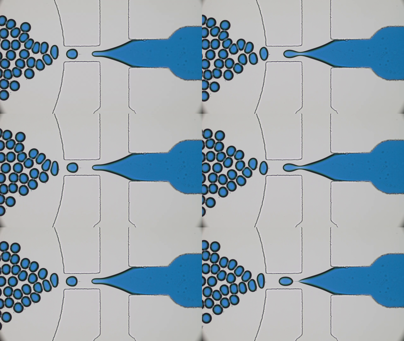
The grand scheme of this research is developing a tool that can selectively shoot drugs into cells at a microscopic level. This is hard because everything happens really slowly at the microscopic scale in a liquid, in ways that meter-sized beings who live in air would not necessarily expect. For example, it is impossible for small organisms to move through a fluid using a repetitive motion that looks the same in reverse. The way we move our feet back and forth to walk would not work for a tiny aquatic human, because the forward motion in the first phase of movement would be nullified by backwards motion in the second phase. This is why bacteria use things like rotating flagella to move*.

Digressions aside, if you tried to shoot a tiny bullet through a cell wall, it would quickly halt and diffuse away before even hitting the cell wall. Soto, Martin, and collaborators wanted to beat this. Perhaps inspired by the likely unrelated Rodrigo Ruiz Soto, a Costa Rican competitive pistol shooter in the 1968 Olympics, Soto sought to develop a cannon that would change the game in the microscopic world in the same way that gunpowder technology changed things in  the macroscopic world.

The researchers developed a “microcannon,” starting with a thin membrane of polycarbonate plastic studded with small pores, which is a thing you can buy and don’t have to make. The pores would eventually serve as the molds for the barrels of the cannons. They deposited graphene oxide onto the inside of the pores using electrochemistry, and then sputtered gold onto the inside of that graphene layer.  While they were still in the plastic membrane, the cannon pores were filled with a gel (literally gelatin from the supermarket) loaded with micron-sized plastic beads to act as bullets, and the “gunpowder,” which I’ll describe after the next image. The polycarbonate is then washed away with acid, leaving free-floating carbon and gold cannon barrels a few microns in size.

cannon
Figure 1: The microcannons, loaded with nanobullets before and after firing. Adapted from Soto and collaborators

While it is generally difficult to make small things move quickly in a fluid, bubbles are somewhat of an exception to this rule. Their collapse can lead to rapid motion on tiny scales. Taking advantage of this, the authors used perfluorocarbon (molecules with the same structure as hydrocarbons but with fluorine connected to carbon atoms instead of hydrogen) droplets as a propellant, which they turned into bubbles with an ultrasound-induced phase transition (essentially blasting them with soundwaves until they vaporized). When they initiated the collapse of the bubbles, they emitted a pressure wave which drove the nanobullets out of the barrel towards their target**.

cannon2
Figure 2: Composition and operation of the microcannons.

The authors performed two tests to characterize how powerful these things were. First, they embedded the cannons in an agar gel (an algae-based substance that Japanese desserts are made of) and loaded them with fluorescent beads. They looked at where the beads were before firing the ultrasound trigger at the cannon, and after. They observed that the beads had penetrated an average of 17 microns through the gel. However, this is about the thickness of a human cell layer, so this could be used, for example, to shoot a small amount of medication through the layer of cells on the wall of a blood vessel. In some more direct studies of the damage caused by collapsing bubbles (which is a common mechanism of damage to ship propellers), the jets that formed when bubbles collapse were shown with high-speed photography to penetrate about a millimeter into a gel. However, these bubbles were 1000 times bigger than those formed in the microcannons, and it’s not out of the question to assume that the penetration depth scales with bubble size.

jjrqrsy
Figure 3: High-speed photography of a millimeter-sized bubble collapsing near a gel wall and shooting a jet into the gel. The mechanism of nanobullet-firing and penetration is a smaller version of this. From Brujan, Emil-Alexandru, et al. “Dynamics of laser-induced cavitation bubbles near an elastic boundary.” Journal of Fluid Mechanics 433 (2001): 251-281.

The bullets were too fast to record with a microscope camera, so their second test involved recording the motion of the cannon after it fired the bullets. Naively, one would expect to be able to calculate the bullet speed with conservation of momentum from knowing the cannon’s speed, but momentum isn’t conserved in a noisy viscous environment (which brings us back to why it’s so hard for microorganisms to move around). They modeled the fluid dynamical forces acting on the system, measured that the terminal speed of the cannon was about 2 meters per second, and concluded that the initial speed of the bullets is 42 meters per second or 150 kilometers per hour (see appendix). Pretty fast, especially for something so small in a draggy environment.

After finding this paper I emailed the first author, Fernando Soto, saying that I enjoyed his paper, and he responded by saying that he was glad that other people liked his “very sci-fi nanodream.” I don’t know if this technology will succeed in the authors’ goal of localized drug delivery to cells, but I think it’s awesome that they made a functioning microscale cannon.

cannon3
Oh the humanity.

*I recommend reading Life at Low Reynolds Number if this interests you.

**Or just in whatever direction it was pointing, I guess.


Appendix: Velocity calculation

The researchers wanted to figure out how fast the bullets were moving based on their measurement of how fast the cannons were moving. Normally you could just use conservation of momentum, but because of the surrounding fluid, momentum is not necessarily conserved (unless you know the momentum of the fluid as well).

However, we understand how velocity decreases in a fluid based on drag: if the velocity is low, the drag force arises from separating the water molecules from each other, and the force is linear with velocity. If the velocity is high, the force arises mainly from accelerating the water to the speed of object, and the force is quadratic with velocity. To figure out which rule applies you can calculate what’s called the Reynold’s number, Re, which is the ratio of inertial to viscous forces in a fluid. If Re is in the thousands or higher,the flow is turbulent. f it’s below 100, the flow is smooth, or laminar. Specifically, the Reynold’s number is calculated as:

$latex Re=\frac{\rho L v}{\mu}$

where $latex \rho$ is the density of the fluid, L is the length of the object in the flow, v is its velocity, and $latex \mu$ is the viscosity. The microcannon was seen moving at about a micron per second, it was about 15 microns long, and the high speed photograph was done in water (density of 1 kg/L, viscosity of about 0.001 pascal seconds). This means the Reynold’s number was about 13, in the laminar regime, and that drag is due to viscosity and linear.

The equation of motion for a slowing object undergoing viscous drag with an initial velocity is

$latex v(t)=v(0)e^{-kt/m}$

where m is the mass of the cannon (known from stoichiometry) and k is the drag coefficient which depends on the viscosity as well as the geometry of the object experiencing drag. Because they know v(t) (as determined from high speed videography), t (the time since detonation), k, and m, they can find v(0).

Then it is assumed that momentum is conserved during the detonation, so the nanobullets with known mass can have their velocity calculated from

$latex v_{c}m{c}=v_{b}m_{b}$

where the indices c and b refer to cannon and bullet. The velocity was calculated to be 42 m/s. Pretty fast.