Putting the controversy over atomic-molecular theory to rest

Original paper: Einstein, Perrin, and the reality of atoms: 1905 revisited


There are many things that we “know” about the world around us. We know that the Earth revolves around the Sun, that gravity makes things fall downward, and that the apparently empty space around us is actually filled with the air that we breathe. We take for granted that these things are true. But how often do we consider whether we have seen evidence that supports these truths instead of trusting our sources of scientific knowledge?

Students in school are taught from an early age that matter is made of atoms and molecules. However, it wasn’t so long ago that this was a controversial belief. In the early 20th century, many scientists thought that atoms and molecules were just fictitious objects. It was only through the theoretical work of Einstein [1] and its experimental confirmation by Perrin [2] in the first decade of the 20th century that the question of the existence of atoms and molecules was put to rest. Today’s paper by Newburgh, Peidle, and Rueckner at Harvard University revisits these momentous developments with a holistic viewpoint that only hindsight can provide. In addition to re-examining Einstein’s theoretical analysis, the researchers also repeat Perrin’s experiments and demonstrate what an impressive feat his measurement was at that time.

In the mid-1800s, the botanist Robert Brown observed that small particles suspended in a liquid bounce around despite being inanimate objects. In an effort to explain this motion, Einstein started his 1905 paper on the motion of particles in a liquid with the assumption that liquids are, in fact, made of molecules. According to his theory, the molecules would move around at a speed determined by the temperature of the liquid: the warmer the liquid, the faster the molecules would move. And if a larger particle were suspended in the liquid, it would be bounced around by the molecules in the liquid.

Einstein knew that a particle moving through a liquid should feel the drag. Anyone who has been in a swimming pool has probably felt this; it is much harder to move through water than through air. The drag should increase with the viscosity, or thickness, of the fluid. Again, this makes sense: it is harder to move something through honey than through water. It is also harder to move a large object through a liquid than a small object, so the drag should increase with the size of the particle.

Assuming that Brownian motion was caused by collisions with molecules, and balancing it with the drag force, Einstein determined an expression for the mean square displacement of a particle suspended in a liquid. This relationship indicates how far a particle moves, on average, from its starting point in a given amount of time. He concluded that it should be given by

$latex \langle \Delta x (\tau) ^2 \rangle = \frac{RT}{3 \pi \eta N_A r} \tau$

where R is the gas constant, T is the temperature, $latex \eta$ is the viscosity of the liquid, $latex N_A$ is Avogadro’s number [3], r is the radius of the suspended particle, and $latex \tau$ is the time between measurements [4]. With this result, Einstein did not claim to have proven that the molecular theory was correct. Instead, he concluded that if someone could experimentally confirm this relationship, it would be a strong argument in favor of the atomistic viewpoint.

A man using a camera lucida to draw a picture of a small statue.
Figure 1: A camera lucida is an optical device allows an observer to simultaneously see an image and drawing surface and is therefore used as a drawing aid. (Source: an illustration from the Scientific American Supplement, January 11, 1879)

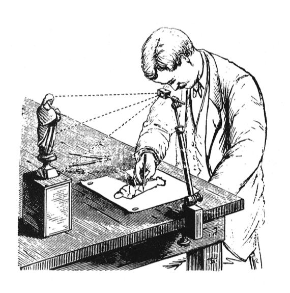
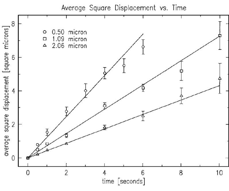
This is where Perrin came in. Nearly five years after Einstein’s paper was published, he successfully measured Avogadro’s number using Einstein’s equation, confirming both the relationship and the molecular theory behind it. However, with the resources available at the time, this experiment was a challenge. Perrin had to first learn how to make micron-size spherical particles that were small enough that their Brownian motion could be observed, but still large enough to see in a microscope. In order to measure the particles’ motion, he used a camera lucida attached to a microscope to see the moving particles on a surface where he could trace their outlines and measure their displacements by hand. Perrin obtained a value of $latex N_A = 7.15 \times 10^{23}$ by measuring the displacements of around 200 distinct particles in this way.

Performing this experiment in the 21st century was much simpler than it was for Perrin. Newburgh, Peidle, and Rueckner were able to purchase polystyrene microspheres of various sizes, eliminating the need to synthesize them. They also used a digital camera to record the particle positions over time instead of tracking the particles by hand. Using particles with radii of 0.50, 1.09, and 2.06 microns, they measured values of $latex 8.2 \times 10^{23}$, $latex 6.4 \times 10^{23}$, and $latex 5.7 \times 10^{23}$. Perhaps surprisingly, even with all of their modern advantages, the researchers’ results are not significantly closer to the actual value of $latex N_A = 6.02 \times 10^{23}$ than Perrin’s was a hundred years earlier.

A plot of the average mean square displacement of three different sized particles over time.
Figure 2: Einstein’s relationship predicts that the mean square displacement should be linear in time. By observing this relationship for three different particle sizes, the researchers use the slope to obtain three measurements of Avogadro’s number. (Newburgh et al., 2006)

For those of us who work in the field of soft matter, the existence of Brownian motion and the linear mean square displacement of a particle undergoing such motion are well-known scientific facts. The authors of this paper remind us that, not so long ago, even the existence of molecules was not generally accepted. And, although we often take for granted that these results are correct, first-hand observations can be useful for developing a deeper understanding and appreciation: “…one never ceases to experience surprise at this result, which seems, as it were, to come out of nowhere: prepare a set of small spheres which are nevertheless huge compared with simple molecules, use a stopwatch and a microscope, and find Avogadro’s number.” [5]


[1] A. Einstein, “On a new determination of molecular dimensions,” doctoral dissertation, University of Zürich, 1905.

[2] J. Perrin, “Brownian movement and molecular reality,” translated by F. Soddy Taylor and Francis, London, 1910. The original paper, “Le Mouvement Brownien et la Réalité Moleculaire” appeared in the Ann. Chimi. Phys. 18 8me Serie, 5–114 1909.

[3] Avogadro’s number is the number of atoms or molecules in one mole of a substance.

[4] In 1908, three years after Einstein’s paper, Langevin also obtained the same result using a Newtonian approach. (P. Langevin, “Sur la Theorie du Mouvement Brownien,” C. R. Acad. Sci. Paris 146, 530–533 1908.)

[5] A. Pais, Subtle Is the Lord (Oxford U. P., New York, 1982), pp. 88–92.

The matter of maternal mucus: permeability and preterm birth

Original paper: Probing the potential of mucus permeability to signify preterm birth risk


What is the first thing that comes to mind when you hear the word mucus? For most people, it’s probably the last time they had a cold. Mucus is not usually something we think about unless there’s a problem. However, it is always there, working behind the scenes to make sure that our bodies function smoothly. Mucus lines the digestive, respiratory, and reproductive systems, covering a surface area of about 400 square meters- about 200 times more area than is covered by skin. In addition to providing lubrication and keeping the underlying tissue hydrated, mucus also plays a key role the human immune system. It serves as a selectively permeable membrane that protects against unwanted pathogens while also helping to support and control the body’s microbiome [1].

Mucus is an example of a hydrogel, which is a three-dimensional polymer network that is able to hold a large amount of water. While hydrogels get their structural integrity from this polymer network, the polymer makes up only a small fraction of the material once they are swollen with water [2]. In mucus, this network is made of biopolymer called mucin.

Researchers in the Ribbeck lab at MIT think that mucus is an underappreciated–and understudied–part of the human body. They have developed techniques for characterizing the mucus hydrogel to better understand how it is able to function as a selective filter. In today’s paper, Kathryn Smith-Dupont and coworkers in the Ribbeck lab investigate cervical mucus and try to understand the relationship between mucus permeability, or its ability to be a selective filter, and the risk of preterm birth.

A birth that occurs before 37 weeks of gestation is considered a preterm birth. This can be associated with negative health outcomes for the baby both in infancy and later in life. Preterm birth is the leading cause of death for children 5 years of age and under, and those who survive can face challenges such as learning disabilities and hearing problems [3]. While the causes of preterm birth can be complex and varied, infection in the fluid surrounding the fetus–which is known to trigger preterm birth–is seen in 25-40% of cases. The infecting bacteria are often the same species that are found in the vagina, suggesting that it traveled through the cervical mucus barrier to infect the sterile uterus.

Smith-Dupont and coworkers look for correlations between mucus permeability and preterm birth risk by comparing the cervical mucus in ovulating non-pregnant women with that in pregnant women. Once the pregnant women give birth, their mucus is characterized as low-risk or high-risk depending on whether they had a preterm birth. The cervical mucus in ovulating non-pregnant women is expected to be at its most permeable to facilitate the passage of sperm, whereas in pregnant women the mucus should be less permeable. Whether a microbe makes it through the mucus barrier can be affected by its size, biochemical interaction with the mucin, or a combination of the two.

First, the researchers look at the permeability of the mucus to 1-micrometer spheres. This is comparable in size to both the mucus mesh and bacteria, and is used to see if the structure of the mucin network is hindering transport through the mucus. Next, they look at the permeability of the mucus to nanometer-size peptides (small bio-molecules). These are much smaller than the mucus mesh, so their ability to pass through the mucus is determined by biochemical interactions with the mucus instead of by its structure. By using these two probe sizes, the researchers hope to identify which mechanism is responsible for any differences in the mucus permeability.

msd_2plots_anno2
Figure 1: (a) Examples of trajectories of particles with ballistic (blue), diffusive (green), and subdiffusive (red) behavior. (b) The MSD for each trajectory on a log-log plot. An MSD with a slope of 2 or 1 indicates ballistic and diffusive behavior, respectively. An MSD with a slope smaller than 1 indicates subdiffusive motion.

To quantify the motion of 1 micrometer spheres in the mucus, the researchers track the motion of spheres in each mucus sample and calculate their mean square displacement (MSD). A particle’s mean square displacement describes how far it moves, on average, from its starting point in a given amount of time. The MSD is characterized by

$latex \langle r \left( t \right) ^2\rangle = 4 D_{\alpha} t^{\alpha}$

where $latex \langle r^2 \left( t \right) \rangle$ is how far the particle is from its starting point after t seconds and $latex D_{\alpha}$ describes how quickly the particle moves (called the diffusion coefficient). If a particle is acted on by a constant force, it moves in a straight line known as ballistic motion and $latex \alpha = 2$. This is not how a micrometer-scale particle in a fluid moves because it is being bounced around by random forces from the molecules in the fluid. Instead of moving in a straight line, the particle’s trajectory is a series of small excursions in random directions, and it takes longer to get away from its starting point than if it just moved in a straight line. This type of motion is known as free diffusion, and its MSD is characterized by $latex \alpha = 1$. In mucus, the polymer network gets in the way of the particle’s diffusion, so it can’t diffuse freely. This motion is called subdiffusive, and it has $latex \alpha < 1$. The more the particle’s diffusion is hindered by the polymer network, the lower its value of $latex \alpha$ will be. An example of a trajectory and MSD plot for each type of motion is shown in Figure 1.

mucus_subdiffusion
Figure 2: The diffusion coefficient $latex D_{\alpha}$ (a) and the diffusion exponent $latex \alpha$ (b) from the single particle tracking of 1 micrometer spheres in mucus samples. (Adapted from Smith-Dupont et al., 2017)

To compare the permeability of the mucus samples, the researchers measure $latex \alpha$ and $latex D_{\alpha}$ for each sample, as shown in Figure 2. The mucus from the pregnant women resulted in lower values of $latex \alpha$ and $latex D_{\alpha}$ than in the non-pregnant women, indicating that the network is more restrictive, as expected. However, the small difference between the high-risk and low-risk pregnancy women was not statistically significant [4]. This suggests that the difference in mucus permeability between high-risk and low-risk pregnancies is not primarily caused by differences in the mucus mesh size.

Next, the researchers look at the permeability of the mucus to small, fluorescently labeled peptides. They use a microfluidic device (to learn more about microfluidics, see [5]) to flow a solution of the peptides through the mucus, and observe whether the peptides get trapped or are able to flow through by looking at the fluorescent profile. Figure 3 shows a schematic of the microfluidic device. The ability of a small particle to travel through mucus is controlled by what happens when it comes in contact with part of the network. This interaction is thought to be affected by the charge of the particle, so the researchers investigate the behavior of both positively and negatively charged peptides.

mucus_microfluidic
Figure 3: A schematic of the microfluidic device used to determine the permeability of mucus samples to fluorescently labeled peptides. If the mucus is not permeable to the peptides they get stuck in the mucus, causing enrichment (or buildup) of peptides at the front of the mucus sample. If the mucus is permeable, the peptides penetrate the mucus and are seen throughout the sample. (Adapted from Smith-Dupont et al., 2017)

For both positively and negatively charged peptides, the researchers see a significant difference between low-risk and high-risk mucus, as shown in Figure 4. The mucus from both low-risk and high-risk patients was less permeable to the positively charged peptides than the mucus from the ovulating patients. However, more of the positively charged peptides were able to penetrate into the high-risk mucus than the low-risk mucus. The results for the negatively charged peptide were more dramatic. While the low-risk mucus was not permeable to the negatively charged peptide, the high-risk mucus was as permeable as that from the ovulating patients. This suggests that the biochemical properties of the cervical mucus in low-risk and high-risk patients are primarily responsible for differences in permeability.

mucus_peptides
Figure 4: Fluorescence profiles after 900 seconds for positively and negatively charged peptides through mucus samples. A control shows the profile in fluid with no mucin. (Adapted from Smith-Dupont et al., 2017)

The results in this study help to clarify which properties of cervical mucus cause an increased risk of preterm birth. The researchers considered both structural and biochemical origins for the increased permeability of cervical mucus to harmful pathogens. Structural changes in the mucin network do not appear to be the primary difference between cervical mucus in low-risk and high-risk pregnancies. Instead, biochemical changes in the mucus that affect how the mucus interacts with microbes appear to be the primary cause of its increased permeability in high-risk pregnancies. This understanding could be useful for developing diagnostic tools to determine a woman’s preterm birth risk and, ideally, treatment to reduce her risk.


[1] https://en.wikipedia.org/wiki/Mucous_membrane#cite_note-Sompayrac-3

[2] Ahmed, Enas M. (2015). Hydrogel: Preparation, characterization, and applications: A review. Journal of Advanced Research, 6(2), 105-121.

[3] http://www.who.int/mediacentre/factsheets/fs363/en/

[4] While the difference between high-risk and low-risk pregnant women is not significantly significant, this does not rule out a difference between the two. The sample size is relatively small for this study, with only 14 pregnant women (7 low-risk and 7 high-risk) included, so the lack of statistical significance could also be due to insufficient data.

[5] https://www.nature.com/articles/nature05058.pdf?origin=ppub

Scientists dream of micro-submarines

Original paper: Graphene-based bimporphs for micron-sized, autonomous origami machines


In the 1966 movie Fantastic Voyage, a submarine and its crew shrink to the size of a microbe in order to travel into the body of an escaped Soviet scientist and remove a blood clot in his brain. The film gave viewers a glimpse into a possible future where doctors could treat patients by going directly to the source of the problem instead of being limited by the inaccessibility of most parts of the human body. This dream of a tiny submarine that can be piloted through the human body to deliver medical care remains, even 50 years later, in the realm of science fiction. However, Miskin and coworkers at Cornell University have brought us one step closer to making this a reality with their recent development of autonomous microscale machines.

To live up to its name, an autonomous machine must have two features. First, it should be able to detect a stimulus from its environment. Then, without any help or intervention, it must respond to the stimulus with a desired response. In this scenario, the machine is not thinking or making decisions— instead, its response to the stimulus is pre-programmed. The ability to respond without supervision means that it can function in remote, inaccessible places, such as deep inside the human body.

One of the biggest challenges to miniaturizing machines is that they contain moving parts. Even fairly simple mechanisms like hinges and valves are too difficult to make on such a small scale. They would require sub-micron machining precision that is not possible using techniques available today. As a result, scientists and engineers must develop alternative mechanisms to perform the functions of these moving parts.

To address this problem, McEuen and Cohen develop a bimorph actuator— a mechanism that allows the machine to move in response to a stimulus, but does not have any complicated moving parts to fabricate [1]. Instead, the bimorph actuator is just a very thin sheet with two layers, one of graphene and the other of glass, that bends in response to changes in temperature or electrolyte concentration. The glass layer expands or contracts when exposed to the different environmental conditions [2], but the graphene does not.  The expansion or contraction of only one of the layers causes the whole sheet to bend (as shown in Movie S2) [3]. Although glass seems like a material that would break instead of bending, the actuator is only two nanometers thick so it bends easily.

nanosubmarines_fig1
Figure 1: The rigid panels on the bimorph sheet direct it to fold into the desired shape. (Adapted from Miskin et al., PNAS 2017)

To harness the motion generated by their bimorph actuator, the researchers take inspiration from an old technique: origami. Since the 17th century, origami has been used in Japan to transform flat sheets of paper into three-dimensional sculptures using only a series of folds. With paper origami, the person making the folds knows where they need to go to make the right final sculpture. However, for a micro-machine, these folding instructions must be programmed into the flat sheet during fabrication so it can fold itself. To do this, the researchers attach thick, rigid panels to certain areas of the bimorph sheet, as shown in Figure 1. The sheet is then only able to fold in the areas between the panels, so the folds are constrained by the shapes and locations of the panels. Using this technique, the researchers construct a variety of structures including a helix, a tetrahedron, a cube, and even a book with clasps, as shown in Figure 2.

nanosubmarines_fig2
Figure 2: Using bimorph actuators, the researchers make complex three-dimensional figures. On the left, the unfolded structure. Center, the folded structures, all shown with the same scale. Right, the same structure folded from paper. (Adapted from Miskin et al., PNAS 2017)

While a self-folding cube is still a long way from a submarine, this technology does open the door to the development of small machines that function on the cellular level. All of the materials used in the origami micro-machines are biocompatible, so they are non-toxic to cells yet robust enough to withstand the conditions inside the body. The closed structures could potentially be used in the body to selectively deploy a drug in response to a local environment.

With further refinement, these machines have the potential to do more complex things. They are strong enough to support electronics and still be able to fold. In fact, the faces of the folded structures are large enough to contain a microprocessor with about 30 megabits of memory or even a functional radio-frequency identification (RFID) chip. The graphene layer in the bimorph also retains its electrical properties, which may allow for the creation of a network of electrically-connected origami machines that can do more complicated tasks than one machine on its own. So, while these origami machines may be simple, they are a step toward precise sensing and manipulation of matter on the cellular scale and—maybe someday—a microscopic submarine.


[1] Bimorph, meaning “two-shape” or “two-form”, refers to the two layers of different materials. In this case, one of the materials responds to changes in the environment to produce bending. In general, either one or both materials can be active. Bimorphs are commonly used for actuation, or generating motion, as shown in this paper. They can also be used for sensing by making one of the materials is piezoelectric so it generates a voltage when it bends.

[2] The ion exchange process is well-known for being able to swell glass and is used commercially to make chemically toughened glass. In certain electrolyte or pH conditions, alkali metal or hydronium ions can diffuse into the voids in the glass and associate with dangling silicon-oxygen bonds. If the ion is larger than the pre-existing void, this causes the glass to swell. Larger ions, such as potassium, result in more swelling than smaller ions like sodium.

[3] This is the same bending mechanism by which a bimetallic strip can be used in a thermostat. The strip, which is made out of two metals that expand differently due to temperature, is made into a coil whose curvature then depends on the temperature and tells the thermostat when to adjust the temperature and in what direction.

Rebuilding hard matter with soft matter

Original paper: Composite Colloidal Gels Made of Bisphosphonate-Functionalized Gelatin and Bioactive Glass Particles for Regeneration of Osteoporotic Bone Defects


The skeleton is the backbone of the body, both literally and figuratively. Healthy bones protect soft organs from injury and enable the body to move. Starting from childhood, staying active and following a healthy diet helps the body maintain healthy bones. However, as people age, their bones can start to weaken. There are often no early symptoms to weakening bones, and as a result the first indication of a problem may be a painful break once the weakening has already significantly progressed.

Although it may seem like bones are made of a hard material, they are actually an elegant combination of hard and soft materials. A primary component of bone is collagen, which forms a soft protein network. This network then provides a scaffold for calcium phosphate, a mineral that provides bone with its hardness and strength. This mixture of hard and soft material enables bone to be flexible enough to withstand impacts, but rigid enough to maintain its structural integrity.

Bone tissue, like other tissue in the body, is alive and therefore able to grow and heal. The material in bones is always being resorbed (or removed) by the body and simultaneously replaced. Around the age of thirty, the rate of resorption overtakes the rate of replacement, causing bones to slowly weaken. When these rates become too disparate, osteoporosis can develop, causing bones to become weak and porous [1].

Bone fractures are more common in patients with osteoporosis, and when they occur, treatment is often needed at the site of the break to promote bone regeneration. The typical treatment in these cases is an autologous bone graft, which involves taking bone from another area in the patient’s body and transferring it to the fracture site to help promote bone regrowth. This technique has two clear downsides: there is limited availability of bone tissue for grafting since it has to come from somewhere else in the person’s body, and removing bone tissue for a bone graft can cause damage to the donor site.

As a result, researchers are working to develop synthetic materials that can provide a better alternative to autologous bone grafts. A promising material would promote bone growth and be strong enough to sustain bending and support weight. Additionally, it needs to be non-toxic and not cause an immune system reaction. Synthetic bone graft materials already exist, but they do not work as well as bone grafts taken from the patient’s own body because they are not as mechanically strong and they do not promote as much bone growth. Hence, the search for better synthetic bone graft materials continues.

In today’s paper, Mani Diba and co-workers investigate a new synthetic material for use in the regeneration of bone tissue in osteoporotic patients. The material in question is a colloidal gel, which is a disordered network structure made of microscale particles suspended in a liquid. This network allows the material to resist applied forces and behave like a solid, even though it may be mostly made up of liquid. Colloidal gels are different from chemically covalent bonded gels [2], like jelly or agar, because their building blocks are microscale particles instead of polymers. These particles bond to each other mostly because they are hydrophobic, or water-repellent, so they would prefer to be next to each other than surrounded by water. The bonds between the colloidal particles are reversible, meaning they can break and reform more easily than covalent bonds in polymer gels, which allows the colloidal gel network to be more adaptable and reform after being broken apart.

The behavior of the colloidal gels is similar to that of toothpaste, which acts like a fluid as it is being forced out of the tube, but once it stops being squeezed it becomes more solid again and doesn’t flow off your toothbrush. For a bone graft material, this means that the colloidal gel can behave as a liquid as it is being injected into a bone defect, and then harden as the network reforms once it’s in place. While this is not a requirement for bone graft materials, it does make the material easier to put in place at a bone defect site.

The researchers prepare a colloidal gel by mixing gelatin particles and glass particles in water (see Figure 1). This choice of particles mimics the structure of bone tissue by using gelatin- a soft material- with glass, which is hard and provides mechanical strength. In order to be a good replacement for a bone graft, the gel must satisfy two main requirements. First, it needs to be mechanically robust to serve as a load-bearing scaffold for bone growth. Second, it needs to be biocompatible, meaning that it should support the growth of new bones.

fig1_all
Figure 1: (a) A schematic showing the formation of a gel by mixing glass and gelatin particles (b) Electron microscopy images of colloidal gelatin (left) and glass (right) particles (adapted from Diba et al.)

The first set of experiments in this paper look at the mechanical properties of the colloidal gel by measuring its storage modulus, which characterizes how strong the gel is. The researchers find that increasing the ratio of glass to gelatin particles or increasing the total number of particles in the gel increases the storage modulus by a factor of more than 100, from about 0.1 kilopascal to tens of kilopascals. The gel is also able to recover its initial storage modulus after being broken apart by shearing, similar to how silly putty can recover its mechanical properties after being stretched. This indicates that the network is able to reform in the bone and become solid again, as expected.  

After characterizing the gel’s mechanical properties, the researchers investigate whether it can promote new bone growth. The growth of new bone starts with the multiplication of osteoblasts, or bone-forming cells, that produce bone matrix material. A signature of this process is an increase in the levels of certain enzymes. Once the matrix is well formed, it undergoes mineralization, which is the deposition of inorganic material (calcium) onto an organic matrix (collagen). This process can be monitored by measuring the amount of calcium added to the area [3]. The researchers track these two indicators, enzyme levels and calcium deposition, to measure the biocompatibility of the gels.

Diba and coworkers study the biocompatibility of the gels both in test tubes and in living animals. In the test tubes, they only find significant mineralization at a glass to gelatin ratio of 0.5 (the highest investigated), which also corresponds to the largest peak in enzyme levels. For testing in animals, the researchers therefore opt for a composite gel with a glass to gelatin ratio of 0.5 and compare the bone growth to that with a single-component gel with no glass particles. The researchers implant these gels in bone defects in the femurs of osteoporotic rats and measure the amount of bone growth after 8 weeks.

Surprisingly, in the rats, the addition of glass particles to the gel did not increase the amount of bone mineralization beyond that seen in the single-component gel as the researchers hoped. However, the bone growth in the composite gel did show more blood vessel-like structures than in the single component gels (see Figure 2), which is important because bone—like other living tissues—needs blood flow to supply oxygen and nutrients, as well as to remove waste products.

fig3_bone_2
Figure 2: Images of bone regrowth from the composite gel in a rat femur. Left: Bone regrowth in a defect that was originally the size of the black circle. Center: Higher magnification image of the small green rectangle on the left. Black arrows point to blood vessel-like structures. Right: Higher magnification of the red rectangle in the center. Red arrows point to cells observed in the center of the original defect. (Adapted from Diba et al.)

Though the researchers in this study did not find the desired increase of bone mineralization in live rats by using a composite gel instead of a single-component gel, they did see other indicators of improvement. Including glass particles increased the storage modulus of the gel, indicating more mechanical strength. They also saw indicators of improved biocompatibility. The bone growth in the composite gel showed an increase in blood vessel-like structures, and they found test tube results which suggested that including the glass particles may still improve mineralization if a higher ratio of glass to gelatin is used. Considering these improvements over a single-component colloidal gel, this composite colloidal gel is a promising development in the search for better bone graft materials.


[1] https://www.bones.nih.gov/health-info/bone/osteoporosis/overview#b

[2] Covalent bonding is the sharing of valence electrons, which are in the outer shell of electrons, between atoms to make a full valence shell. Any time two non-metals come together they will share their valence electrons.

[3] http://www.promocell.com/fileadmin/knowledgebase/pdf-xls/Osteoblast_Differentiation_and_Mineralization.pdf