Sticky light switches: Should I stay or should I go?

Original paper: Adhesion of Chlamydomonas microalgae to surfaces is switchable by light


One day it’s fine and next it’s…” red? Microscopic algae depend on photosynthesis, so they follow the light. Previous research has shown that their swimming is directed towards white light but not to red light. New work shows that light-activated stickiness allows microscopic algae to switch between different movement methods.
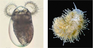
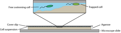
This indecision’s buggin’ me” – should I stick or should I swim? Different types of motility are needed to move through different environments. Microscopic algae live in a variety of different conditions, including soils, rocks, and sands, all surrounded by water. In general, we can split these conditions into two groups: those where the algae move within the water, or those where the algae move across a surface. Today’s paper studies how a unicellular algae changes from its free swimming state to a surface attached gliding state.

swimglide2008.png
Figure 1: Left: Chlamy’s normal swimming beat pattern, with different colors showing different time points. The cell body is shown in blue and the eyespot in red. Image adapted from [1]. Right: Gliding Chlamy moves due to proteins moving within the flagella. Image adapted from [2].
Kreis and co-workers investigate the unicellular green algae called Chlamydamonas reinhardtii, or Chlamy for short. It has two whip-like arms, called flagella, that it uses to move. In the swimming state, the flagella beat in a breaststroke to pull the cell forward, as shown in Figure 1A. In the gliding state, the flagella are stuck to a surface and the transport of proteins inside each flagellum pulls on the surface so the Chlammy moves across the surface, as shown in Figure 1B.

micropipetteFM2008
Figure 2: In micropipette force microscopy a small glass tube holds the cell. A surface (the substrate) can then be moved towards or away from the cell. The deflection of the micropipette as this occurs determines how sticky the cell is. All of this is done in water, where Chlamy lives normally. Image adapted from Kreis and coworkers’ paper.

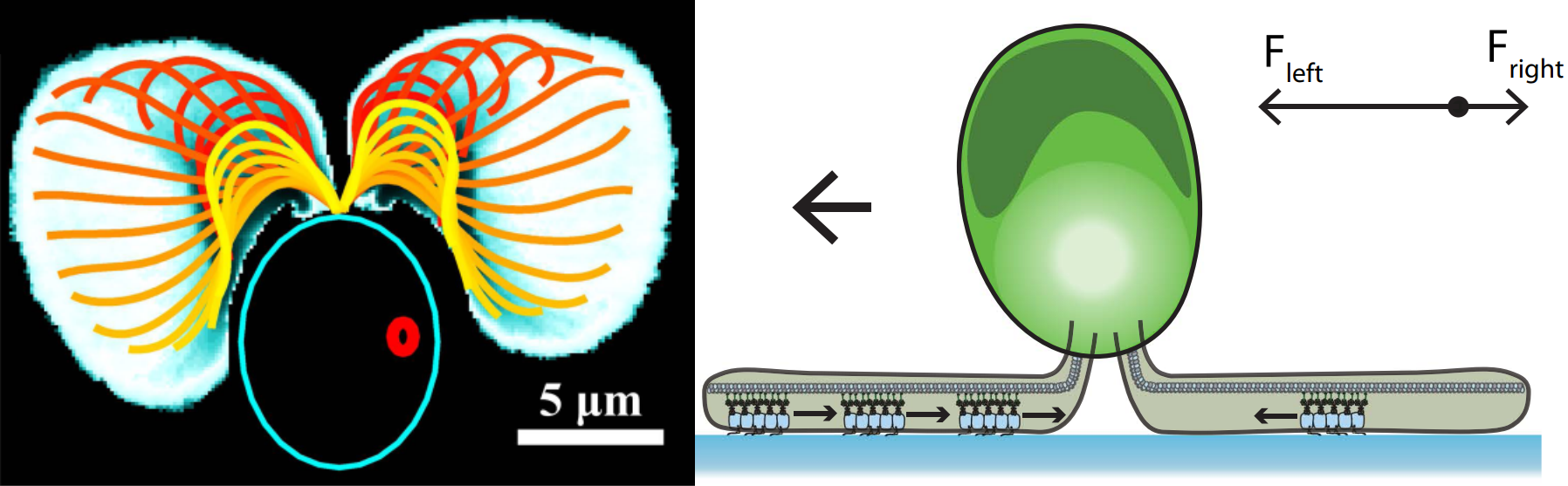
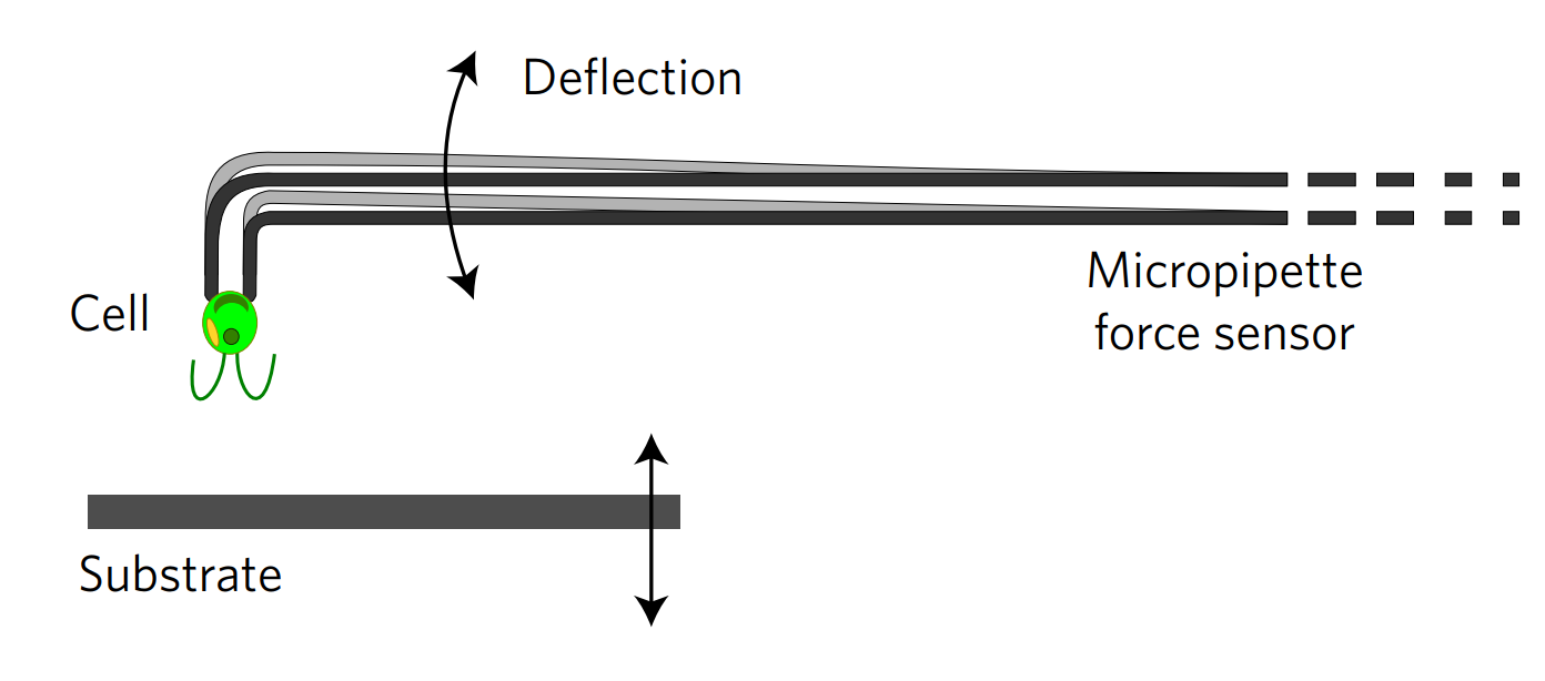
To transition between these two movement methods, the Chlamy must attach and detach from the surface. The researchers measure the force Chlamy exerts on a surface when it attaches using micropipette force microscopy, shown in Figure 2. This method uses a micropipette, which is a small glass tube, to hold a single Chlamy cell in place with suction. The surface is moved towards or away from the cell, deflecting the micropipette from its original position based on the force the cells exert on the surface. The relationship between deflection distance and force is measured beforehand with calibration experiments. So, during the experiment, micropipette deflection yields how strongly cells are stuck. To understand how this force relates to the two movements methods, let’s look at the results.

frontandback2008
Figure 3: Adhesion force as a function of distance from the surface to the cell. The surface is initially 20 micrometers away from the cell and is moved closer, so the cell and surface touch. As the surface is moved away again we can see if the flagella-facing cell (a) or the back-facing cell (b) attach to the surface from the adhesion force that is built up. Figure adapted from Kreis and coworkers’ paper.

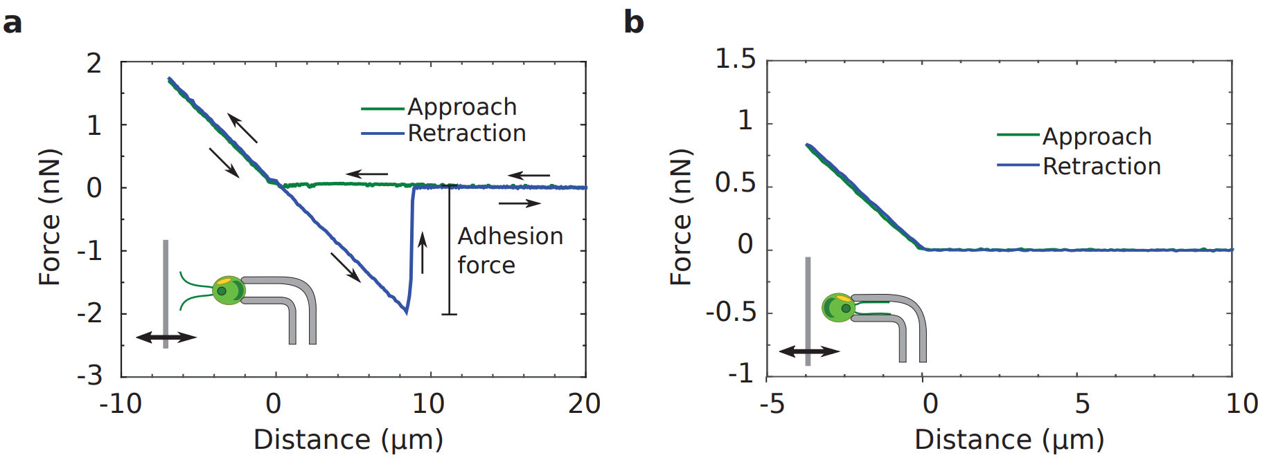
Figure 3 shows two force measurements, one where the flagella are facing the surface and another where the back of the cell is facing the surface. When the surface touches the flagella or back of the cell body, the micropipette is first deflected upwards, giving a positive force. As the surface is moved away, the micropipette moves back to its original zero-force position.

As the surface is moved further away, the flagella-facing cell and back-facing cell behave differently. The flagella-facing cell deflects the micropipette downwards, shown by the build-up of a largely negative force, whereas the back-facing cell does not deflect the micropipette and no force is exerted. This means that the flagella-facing cell sticks to the surface, whereas the back facing cell does not stick.

pullandgraph1009
Figure 4: Top row – left to right shows successive images of Chlamy pulling itself towards a surface – dashed red line shows the movement of the micropipette. The flagella are marked by solid red lines. Bottom row – micropipette deflection over time as the light is turned on and off as indicated by the arrows. Figure adapted from Kreis and coworkers’ paper.

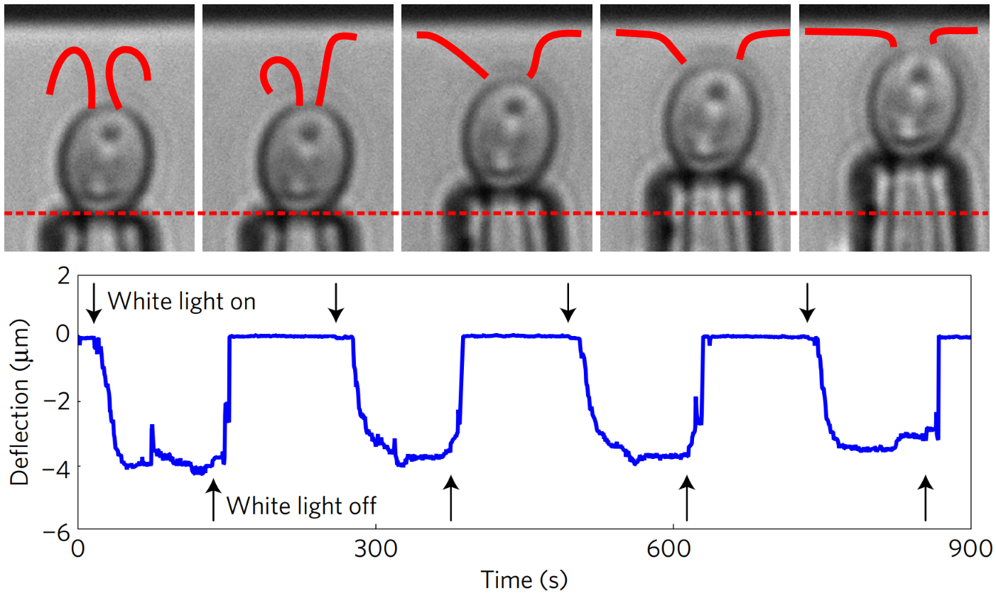
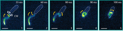
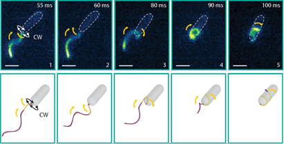
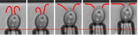
The flagella not only stick but actively pull themselves towards the surface. At the top of Figure 4, we see the flagella touch the surface during their swimming beat cycle. First, just a small part of one flagellum is stuck to the surface. Then, the flagella actively pull themselves towards the surface until both are completely stretched out and ready for gliding. This process is reversible: as the light is turned on and off, so is the adhesion force. The Chlamy can pull themselves up again and again – transitioning between their stuck and free state.

difflight2008
Figure 5: Force-distance curves for the retraction of a surface under different wavelengths of light. The flagella only stick when shorter wavelengths of light are present. Figure adapted from Kreis and coworkers’ paper.

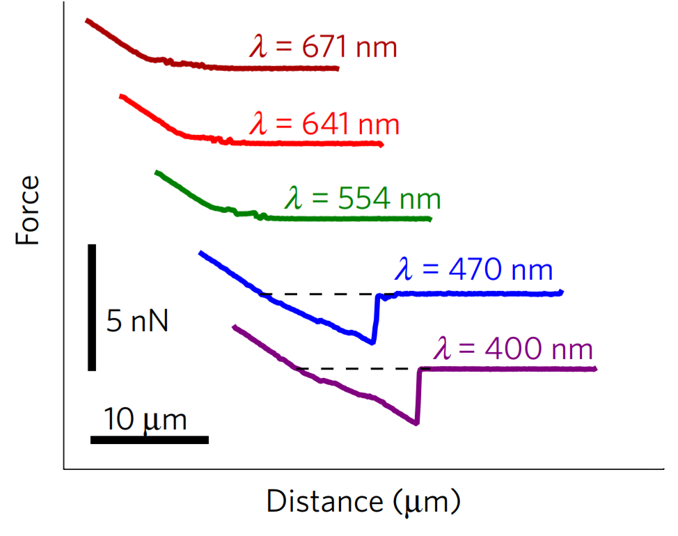
But what controls the transition? To answer this, the researchers repeated the experiment under different wavelengths of light. In Figure 5, we see that the stickiness peak is absent for red and green light but present for blue and purple light. Two potential light sensors could be responsible. One is on the cell’s eyespot and controls cell swimming to guide the cell towards the light. The other is on the flagella and controls the cell life cycle and several aspects of the cell’s mating process. But we don’t yet know which light sensor controls the stickiness, or which specific proteins make the flagella sticky.

So for the Chlamy, the decision to stay or go is made by checking if the lights are on! If they ‘go’ they can seek lighter environments, and if they ‘stay’ they can bask in the sunny spot. Watching Chlamy cells stick and un-stick as we flick a light switch is very cool, but why should we care about Chlamy? Chlamy is used in bioreactors to create biofuels and other bioproducts. Stuck Chlamy prevents light and nutrients from getting to all the cells in the reactor, so we need to understand how to control the sticking process. Plus – if we understand how a simple unicellular organism solves the problems of life, we can use this bio-inspiration for new technologies – in this case possibly new light-switchable adhesives.


[0] Should I Stay or Should I go?

[1] Antiphase Synchronization in a Flagellar-Dominance Mutant of Chlamydomonas

[2] Intraflagellar transport drives flagellar surface motility

EuroScience Open Forum 2018

“ESOF (EuroScience Open Forum) is the largest interdisciplinary science meeting in Europe. It is dedicated to scientific research and innovation and offers a unique framework for interaction and debate for scientists, innovators, policy makers, business people and the general public.” [1]

This year ESOF was in Toulouse, and I was fortunate enough to be able to attend, so I want to share a few snippets of my time there, and my main takeaways.

There was a huge range of different topics on their programme from science communication and careers, to atomic clocks and plastic pollution. I found choosing between parallel sessions was often difficult. But even though I went to a variety of different sessions, I am going to focus on one major theme kept cropping up. Namely, trust.

How can we, as scientists, build trust in science? How can we trust one another?

For this science and scientists need to be seen as credible. There are no quick fixes, but openness was touted by many at ESOF as a huge step towards building more trust. After all if ‘science is not finished until it’s communicated’ [2] then the public are a huge part of science! Not to mention that for most of us, we are in fact paid by taxpayers.  

Here are some of the different types of openness that people discussed at ESOF2018:

  1. Open Access

Currently, many scientific journal articles are behind paywalls – this means institutions without access to a particular journal are locked out. Even when a journal allows institutes to post papers open access from the institute, this is often after long embargo periods restricting access to the latest science.

  1. Open Data

Even if a paper is open access. The methods and the data shown are often too little for experiments or analysis to be easily reproduced. For private and sensitive data – this is not possible but we can strive for the data to be as open as possible and only unavailable when necessary. If the data is accessible and readable, then anyone can reproduce their analysis, particularly if codes are also made accessible and usable.

  1. Open Communication

To share our research with the public, making something understandable to people outside your field is not enough. We need to open a dialogue between scientists and the public so that the outreach activities are catered to their interests. In particular for influencing policy, the people who our science affects need to be heard.

  1. Open Science

Science is a part of our culture, our history and our future. It would be difficult to find anyone who isn’t affected by science. Allowing people to take part is great for building trust. New initiatives and new technologies are opening the ‘ivory tower’ of academia. Citizen science or crowd-sourced science can bring together the public and scientists, for mass collection of data. And new make-spaces, fab-labs and open source software are making it easier for people to conduct their own experiments, build their own devices and explore science in their free time.

A bit of a culture shift needs to occur within science to highlight these aspects of science. I think, most scientists agree openness is important but often these ‘extra’ activities get put on the back burner as publications, teaching responsibilities and funding applications dominate our time. So, more time needs to be invested in openness, but not at the expense of an individual scientist’s career (or home life). Hiring practices, funders and fellow scientists need to reward and encourage openness within science. So I think it is great that so many people from a range of different places were talking about how to increase openness. 

Overall I thought ESOF2018 was a very friendly conference, with so many passionate people working towards a better scientific process. To hear them talk so passionately, either in how science is funded, publishing, science policy, collaborations with industry, scientific careers, science communication and even the science of science communication was a fantastic experience. 

Let us know if you have any experiences, thoughts, or difficulties on accessing science or how to open up science over twitter (@softbites17 or @emilyeriley) or in the comments down below.

[1] https://www.esof.eu/en/

[2] Quote from Sir Mark Walport, who was Chief Scientific Adviser to the UK government.


Imagine you are a Sea Slug Larva…

Original paper: Individual-based model of larval transport to coral reefs in turbulent, wave-driven flow: behavioral responses to dissolved settlement inducer


Lost, alone, and buffeted by ocean currents: this is the beginning of life for many oceanic larvae. These tiny organisms, often only 100 micrometers in diameter, must seek a suitable new habitat by searching over length scales thousands of times their own. But searching for something you can’t see while being dragged this way and that by ocean currents can’t be easy. How do these microscopic creatures make sense of the turbulent world around them and find their home?

larvaandslug
Figure 1: The larval form (left) is about 100 micrometers in diameter and swims using the beating hairs on the stumps on at the top of the cell, whereas and the adult sea slug form (right) can grow up to 5 cm in length and stays on the coral. Left figure is taken from Koehl et. al. The right figure is taken from http://www.seaslugforum.net/find/pheslugu.

To answer this question, today’s paper studies a species of sea slug, Phestilla sibogae. These sea slugs have two forms, the baby larval form (Fig. 1 left), which travels through the ocean, and the adult sea slug form (Fig. 1 right), which lives and feeds on their coral prey. After they are born, the young larvae first swim toward light, instinctively leaving their parents’ reef. When they are old enough to settle down and become adults, they must search for a new reef to call home. The metamorphosis from larva to slug is only triggered when the larvae have settled on their coral prey.

The coral prey release a chemical that the sea slug larvae can smell. The chemical acts as an on-off switch for the larva. When there is no chemical, the larva swims in a straight path in a random direction at 170 micrometers per second. Upon encountering a strong enough chemical smell, the larva stops swimming and sinks at 130 micrometers per second. We know how the larva move but how does this movement affect how many and how quickly the larvae make it to the reef? To understand the larvae transport, we need to understand the larval environment.

larvaeinsea
Figure 2: Larva cell (inset) in turbulent waters above the reef. The streaky pattern is from measurements made by the researchers in a wavy flow tank above a reef skeleton. Within the reef skeleton, a fluorescent dye is released. When the fluorescent dye is excited by a laser sheet it emits light. More light means more dye, where the dye represents the coral chemical [1]. The figure is taken from Koehl et. al.
Not only do the larvae have to swim while being buffeted by the wavy turbulent flow, but the waves also affect how the chemical released from the reef spreads. If the coral were in still water, then the amount of chemical would increase smoothly as you travel from the surface waters to the reef due to diffusion. However, the corals live in shallow water, where waves passing over the rough reef surface lead to turbulent waters above the reef. This complicated flow pattern means the chemical smell no longer smoothly increases as you travel towards to reef. Instead, the turbulence creates streaks of very high amounts of chemical and very low amounts of the chemical, as shown in Figure 2.

To investigate the transport of larvae to the reef, Koehl and coauthors build on previous work to create a computer simulation with both the larva swimming behavior and the larva environment based on experimental measurements. To model the background flow environment, they include the net flow, waves, and turbulence. The flow parameters are fit to experimental measurements made in wavy shallow waters in Hawaii [1]. In a similar way, the researchers use experimental measurements to model the swimming behavior of the larvae.

In their simulation, the researchers are able to alter both the environment and the larva swimming behavior and ask what, if any, advantage the on-off swimming behavior brings. The advantage is measured using the steady state larva transport rate, defined as the percentage of the larvae that make it to the reef each minute. With the steady-state approximation, the percentage of larvae that make it to the reef each minute is constant over time.

bothconcpatterns
Figure 3: Turbulent concentration pattern in A shows streaks of high and low concentration while the time-averaged concentration pattern in B smoothly increases towards the bottom of the reef. Figure adapted from Koehl et. al.

First, the researchers investigate whether or not the streakiness of the concentration pattern is an important factor in determining how many larvae reach the coral. When trying to understand how larvae reach the coral, previous researchers made the simplifying assumption that the concentration pattern of the chemical the larvae follow is smooth and uniform over time. As we saw in the streaky pattern in Figure 2, this is not a realistic assumption. But just how wrong is this it? To answer this question, the researchers compare the chemical distribution measured at a single moment in time (Fig. 3A) to the chemical distribution obtained by taking the average of the distribution measured at many different times. This averaging process produces a smoother distribution than would be seen in reality (Fig. 3B). On comparing the two different chemical distributions, the researchers find the larvae transport rates are overestimated by up to 10% in the unrealistic time-averaged environment.

Secondly, because the concentration pattern affects the transport rate, the on-off swimming behavior must affect the transport of the larvae to the reef. In their simulation, the transport rate for naturally swimming and sinking larvae is 45% per minute. The researchers test how the larva behavior affects this transport rate by separately turning off the swimming and sinking behavior of their simulated larva. If a larva sinks but does not swim, the transport rate changes to 20% per minute. If the larva swims but doesn’t sink, the transport rate changes to 25% per minute. Without their on-off switch, the larvae are reliant on the background flow or randomly swimming downwards to be transported to the coral.

From these transport rates, we can understand the relative importance of larval behavior and larval environment. For example, we now know that if the environment was no longer turbulent or if the larvae could no longer swim, the larvae’s rate of transport to the reef would change significantly. This impacts both how many larvae survive to adulthood and where in the ocean the adult sea slugs end up. Building on this work, predictions have also been made for many different species of larvae [2]. From these studies, we not only can get an idea of how local and global populations spread in their natural environments but also how a simple on-off process can help an organism to successfully navigate a complex environment.


[1] See https://academic.oup.com/icb/article/50/4/539/652640, for an overview of how researchers characterize the larva environment.

[2] See https://link.springer.com/article/10.1007%2Fs00227-015-2713-x for more details. Here, the researchers measure both the concentration of chemical and the flow above the reef simultaneously (as described in [1]). With this, they look more generally at the problem of settling on surfaces, investigating a variety of swimming properties and settling sites rather than a specific species.

Coil and Recoil: New screw-like bacteria swimming

Original Article: Bacteria exploit a polymorphic instability of the flagellar filament to escape from traps

No one likes being stuck. Whether you are in a car stranded in mud or stuck in a dead-end job, continuing normal behaviour is unlikely to help. Whereas we can see approaching hazards and dead-ends and try to avoid them, bacteria must blindly swim through passageways and channels that are of a similar size to themselves, often resulting in the cell becoming trapped. So, how does a bacterium change its behaviour to free itself?

In today’s paper, Kuhn and colleagues report a new type of bacterial motion used to escape from microscopic traps. Like the car stuck in slippery mud, forward-reverse strategies do not free the cell. The bacteria adopt a new swimming setup wherein their swimming appendage is wrapped around the cell body. This new coiled swimming was only discovered in 2017 and currently has only been observed for two bacteria species [1].

For bacteria, which are only a couple of micrometres, the viscosity of the swimming media dominates over any inertial effects. When we swim in water inertia dominates, so we can move to some extent with only one propelling action. However, when there is no inertia, if you stop propelling, you will immediately stop moving. To overcome the limits of their drag dominated environment, many bacteria use thin appendages called flagella.

Bacteria flagella are the only natural structures that generate propulsion via rotary motors. Much like a boat propeller, the bacterial flagellum has a rotary motor at its base, embedded in the cell body that rotates its propeller 100 times a second. However, the bacterial propeller is different from propellers we recognise. The bacteria flagella has two components: the flagellar filament and the hook. The flagellar filament is a helix, 1-3 times the length of the bacterium cell body, and the hook is a nanometre scale elastic segment connecting the filament to the rotary motor. Like a corkscrew, the rotation of the helix creates motion along the helix axis. Both the handedness of the helix, that is which direction the helix curls around its axis, and the direction of rotation determine whether a corkscrew goes forwards or backwards. Typically the bacterial flagellar filament is left-handed and rotates counterclockwise, looking from the end of the flagellum to the cell body, thus pushing the bacterium cell first at about 20 cell body lengths per second. If the same left-handed helix rotated clockwise, the bacterium would swim backwards – filament first – towing the cell body.

F1.largesoftbites
Fig. 1: The experimental setup. The bacteria swimming in a small microstructured environment created by the uneven gel surface. The bacteria rotates its helical propeller counterclockwise transitioning from a free swimming cell to a trapped cell as the bumpy surface come closer to the above thin glass slide. Figure adapted from Kuhn et. al.

To recreate a bacterium’s natural environment, Kuhn and colleagues cover an uneven gel surface by a thin film of liquid with a thin glass slide above, as shown in Figure 1. The varying distance between the bumpy gel surface and the thin glass slide provided a micro-structured environment in which the bacteria could be observed. Their study focuses on a genetically modified soil bacterium that has a single flagellum placed at the end of a pill-shaped body a couple of micrometres in length. By watching the rotation and position of the fluorescently stained 20-nanometre-thick flagellar filament, they are able to determine when the bacterium is stuck and how structural changes enable its escape.

F2.largesoftbites
Fig 2: Approach, trapping and escape. Top row: the bacterium becomes trapped during regular forward swimming. Middle row: backwards swimming is not able to free the cell. Bottom row: the flagellar filament wraps around the cell body and the bacterium is able to reverse to escape the trap. Figure adapted from Kuhn et. al.

Compared to the free-swimming cell in a bulk solution, swimming close to surfaces increases the drag on the bacterium. The cell is trapped when the surfaces are close enough to increase the drag on the cell head above the thrust. To attempt to free themselves, the bacterium switches between clockwise and counterclockwise flagellar rotation – a forward-reverse strategy. This is unsuccessful. A successful escape only occurs when the flagellar filament wraps itself around the cell body, creating a conformation that has not been seen before. The wrapped filament is still helical, so rotation of the motor still creates propulsive forces along the helix axis. The cell then swims in a screw-like motion to release itself, as seen in Figure 2. It is yet unclear why exactly the coiled state enables the bacterium to escape. It could be the change in helical structure, the proximity of the flagellar filament to the cell body or the interaction of the flagellar filament with the nearby surface or even a combination of these fluid drag effects. Once the cell is free, the filament returns to its non-coiled state and normal swimming resumes. But how does this stiff inactive filament change its shape so drastically?

F5.largesoftbites
Fig 3: Flagellar filament transformation. The flagellar filament changes shape sequentially from the base of the helix to the top. The top row shows the experimental images with scale bar 1µm, and the bottom row shows computer simulations with the two different colours of the flagellar filament relating to the two different atomic configurations observed. Figure adapted from Kuhn et. al.

To induce the new swimming state in a bulk fluid environment, the researchers study the bacterium freely swimming in solutions of increasing viscosity. The increased viscosity increases the force on the flagellum and was observed to increase the likelihood of the screw-like motion. The increased forces on the flagellar filament thus seem to be responsible for the drastic change in state. Changes in the helical structure are well known for other flagellated bacteria changing the diameter and even the handedness of the helix, but never as extreme as to wrap the flagellum around the cell. These well-known flagellum changes are due to a change in the atomic structure of the flagellum, so rather than bend elastically, the filament changes shape sequentially along the flagellar filament. As the researchers’ bacterium’s filament is constructed similarly, they ascribe their two swimming types to two atomic configurations available to the flagellum: one normal and one coiled, as shown in Figure 3. Although the coiled swimmer is much slower than the uncoiled swimmer when freely swimming, the coiled state is able to free the bacterium from micro-structured traps, giving the bacterium a significant advantage.

Understanding this new type [1] of bacterial motion is critical to know how a bacterium survives in its natural habitat. For which habitats is the screw motion most common? Are there many more species that can coil to recoil? Where does this screw motion fail? What are the key features of the wrapped state that allow the bacterium to escape? Not only will studying these features help to understand populations of bacteria, for example how they spread and find better habitats, but it could also help in the design of micro-robots within complex environments such as for targeted drug delivery in the body.


[1] The same wrapped swimming configuration has also very recently been observed for a multi-flagellated bacterium, P. putida [you can see the paper here]. This bacterium has 5-7 flagella positioned at one end of the cell. Contrary to the results described above, the transition was observed for cells swimming in a bulk fluid environment. In the coiled state, all 5-7 flagella wrap around the cell. Through modelling the different swimming states of the cell, Hintsche and colleagues show an increased diffusion of populations that are able to transition to a coiled state.