The key to fighting cancer: be flexible

Original paper: Nanoparticle elasticity directs tumor uptake 


In my previous post on soft nanoparticles, you were introduced to polymer-based nanoparticles that could be used in biomedical applications, one of which is cancer therapy. These nanoparticles have a range of useful properties for cancer treatments, including their spherical shape and small size (~100 nm), both of which are similar to exosomes, small globules that are used in nature for transferring proteins between cells. Since cells naturally absorb exosomes, artificial particles with this size and shape should also be easy for cells to absorb, which means these particles could be used to deliver drugs into cells. While this idea sounds promising, it hasn’t worked out in practice —  when drug-loaded polymer-based nanoparticles were injected into a tumor, subsequent tests showed that less than 1% of the injected dose entered the cancer cells. Since these particles were the correct size and shape, why didn’t they get inside the target cells?

One possibility is that the elasticity (or stiffness) of nanoparticles is to blame: scientists have suspected that this mechanical property can affect the ability of nanoparticles to squeeze themselves through the cell’s membrane. Unfortunately, it is difficult to test this hypothesis directly, because modifying the elastic properties of a nanoparticle generally requires modifying its chemical properties as well. To solve this problem, Peng Guo and coworkers designed a special kind of nano-objects — spherical nanolipogels — with tunable elasticity. In this paper, they proved for the first time that breast cancer cells take up soft, squishy particles more easily than they take up hard ones.  

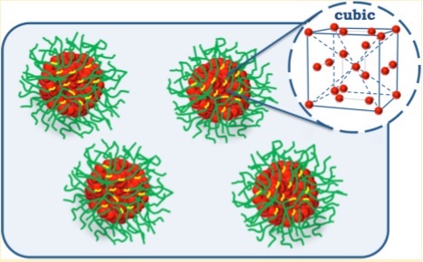
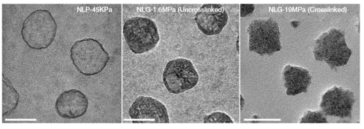
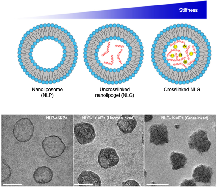
So what are nanolipogels? This type of nanoparticles is basically an altered version of a nanoliposome, a particle-like object that consists of a liquid water core surrounded by a layer of phospholipid molecules [1]. Guo and his colleagues created nanolipogels by filling the nanoliposomes’ liquid core with a polymer of tunable chemical structure. Nanolipogels have precise size (160 nm) and shape (spherical), and their elasticity can be made to vary without changing their other properties (see Figure 1).

image 1
Figure 1. Structures (top) and micrographs (bottom) of nanoliposomes and nanolipogels of increasing stiffness (higher values of Young’s modulus). (Image adapted from Guo’s paper.)
figure 2
Figure 2. Experimental setup of an Atomic Force Microscope. The height of a sample’s surface is scanned by a tip on a moving cantilever and the cantilever deflections are detected by a laser light to give the samples topographic profile. (Image from simple.wikipedia.org)

To measure the elasticity of the particles they had produced, Guo and coworkers used a technique called Atomic Force Microscopy (AFM). AFM is commonly used to visualize soft materials by imaging the height of their surface through the deflection of a cantilever (Figure 2). In this paper, the researchers used AFM for a different purpose: to calculate the Young’s modulus — a measure of stiffness — of the nanoparticles. They did this by compressing the particles between the cantilever tip and a solid surface, allowing the researchers to measure the force required to deform the particles by some known amount. The relationship between the applied force, the degree of deformation, and the Young’s modulus is given by the Hertz equation [2]. What you need to remember is that the greater the modulus, the stiffer the particle.

The researchers created four different nanolipogels of different elasticity with Young’s moduli ranging from 1.6 MPa (roughly the stiffness of cork) to 19 MPa (the stiffness of leather), and a nanoliposome without polymer in the core with a Young’s modulus at 0.045 MPa (roughly the stiffness of gummy bears). After verifying that all 5 particles could successfully encapsulate drug molecules, they tested how well tumor cells could uptake each particle. To do so, they used breast cancer cells in the lab (in vitro cellular uptake) and attached fluorescent dye to the particles to determine whether they were inside or outside of the cells. They found that the stiffest nanolipogels were 80% less effective compared to the softest nanoliposome samples; in other words, five times more of the softer particles got inside the cells. In vivo tumor uptake studies, using live mice, similarly showed that the nanoliposomes had up to 2.6 times higher cellular uptake than the stiffest nanolipogels.

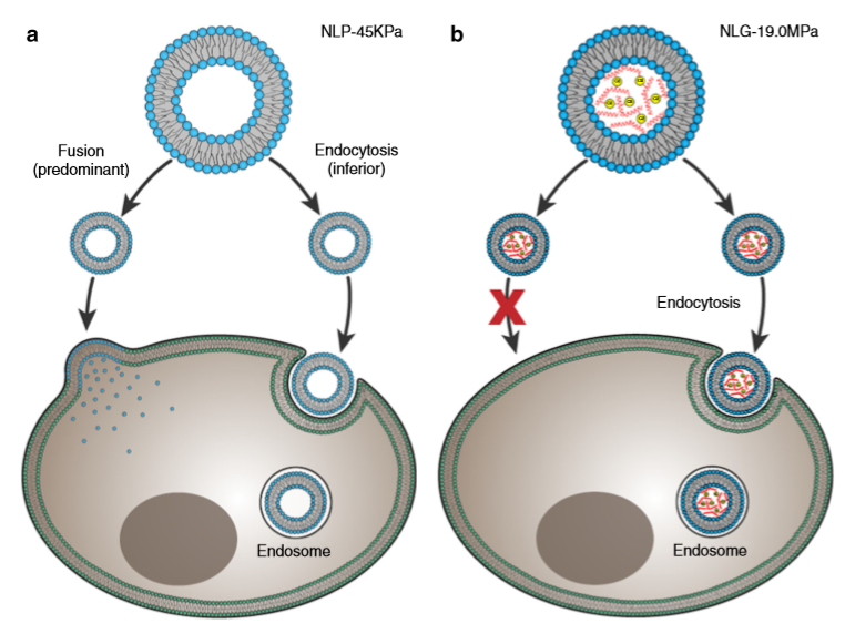
Why do the soft nanoliposomes enter the cells more easily? To understand the conclusion of Guo and colleagues, we need to think about how nano-objects enter a cell. Figure 3 shows two possible ways of doing this: 1. fusion, where nano-objects break up and join the cell membrane, or 2. endocytosis, where the whole object enters the cell by bending the cell’s membrane and getting covered in a membrane outer layer. Fusion needs less energy compared to endocytosis, where cell membrane bending and surface tension increase the energy. The researchers hypothesized that nanoliposomes use both fusion and endocytosis, with a preference for fusion (Figure 3a), while nanolipogels can only enter the cell through endocytosis (Figure 3b). This hypothesis was verified by using chemical compounds that prevented endocytosis from taking place; in all experiments, the cellular uptake of nanoliposomes was as high as before, while much fewer nanolipogels were detected in the cells, since they couldn’t enter through endocytosis.

image 3
Figure 3. The possible pathways of (a) nanoliposomes and (b) nanolipogels entering a cell. (Image adapted by the Guo paper.)

This study showed that a nanoparticle’s mechanical property, in particular, its elasticity, affects how it enters cells, a finding that could potentially have a tremendous impact on cancer treatment and diagnosis. The use of nanoliposomes, which are a synthetic equivalent of nature’s drug delivery systems, may also be used in the future to further understand how cellular processes, such as fusion and endocytosis, take place.


Continue reading “The key to fighting cancer: be flexible”

Anti-biofilm Material to Fight Bacterial Formation on Surfaces

Original paper: Sodium Dodecyl Sulfate (SDS)-Loaded Nanoporous Polymer as Anti-Biofilm Surface Coating Material  


Are you afraid of visiting the dentist? If so, you’re probably not the only one, but unfortunately we can’t avoid it. Yearly dental check-ups are necessary to prevent tooth and gum infections. Dentists use a disturbingly sharp, noisy tool to remove dental plaque from the tooth surface. Dental plaque is caused by bacteria, and it is an example of a biofilm, which is a community of bacterial cells that stick to surfaces. Biofilms can be found everywhere, especially on wet surfaces. Biofilms cause health problems for millions of people worldwide every year, primarily because of infections during surgery or consumption of contaminated packaged foods. To prevent these problems, some scientists are developing surface coatings that will prevent biofilm formation in the first place. In this week’s paper, we will learn about a new technique for creating a microscopic “shield” against the formation and growth of biofilms.

A biofilm is a complicated microscopic world in which multiple bacterial species can coexist. Most parts of the biofilm are covered by a protective sticky slime that is produced by the bacteria themselves. It is now known that bacteria communicate with each other within the biofilm by exchanging small molecules, proteins, genes, and even electrical signals. This intercellular communication results in expression of specific genes throughout the bacterial community in response to the environment. As a result, bacteria in biofilms are able to quickly develop resistance to antibiotics, making the treatment of biofilm infections extremely challenging. Therefore, one of the most common ways of destroying biofilms is a mechanical removal by scraping. This explains why we can’t avoid going to the dentist at least once per year; a toothbrush is not strong enough to remove the biofilm layer that we know as dental plaque.

E.coli-colony-growth.gif
Figure 1. A culture of E. coli growing. Video courtesy of en.wikipedia.org.

Unfortunately, scraping is not always possible; especially in cases where biofilms are formed at surfaces inside the body or on tiny surgical and industrial tools. Li Li and co-workers from the Technical University of Denmark, in collaboration with the Nanyang Technological University in Singapore, developed a coating material with the ultimate goal of preventing the growth of multiple bacterial species. This material has a structure with many nanoscale pores (holes) able to load and release antimicrobial compounds. For this study, it was filled with a detergent compound that is part of household cleaning products and is known to kill bacteria by dissolving the bacterial cell membrane. The researchers loaded the nanoporous polymer films with the detergent and placed the films in contact with Escherichia coli (E. coli) biofilms. Before showing what happened to the E. coli biofilms, let’s discuss a little bit more about the nanoporous polymer film.

figure 1 gyroid
Figure 2. The periodic gyroid structure of the nanoporous polymer films. Video courtesy of en.wikipedia.org.

The nanoporous film used in this study is made of a polymer that has two hydrophobic (water-repelling) chains, one of which is a silicon-based material that we use in contact lenses. The polymer chains self-organize into the beautiful gyroid structure shown in Figure 2, which is a 3D interconnected surface that repeats in three directions (is triply periodic) and contains no straight lines. The most beneficial part of this structure is that it forms small, nanoscale pores after the removal of the silicon-based chains, which provide large storage space for the detergent molecules. To stabilize the final structure used in this study, the researchers add a chemical compound to remove the silicon-based chains. At this step, the interconnected polymer chains form strong covalent bonds with each other, a process called cross-linking. Figure 3 shows the process of the nanoporous film preparation (a, b) and loading of the detergent (c, d) (to learn more about the preparation, see [1]).

figure 2 nanoporous film
Figure 3. Representation of making the nanoporous polymer film and loading it with detergent by diffusion: (a) the block copolymer re-organizes into a gyroid structure, (b) the silicon-based polymer chains are removed from the nanoporous film, (c) The detergent solution is in contact with the nanoporous film and the detergent molecules attach to the pore walls (the enlargement shows that excess free detergent molecules may form small spheres between the walls), (d) the final nanoporous film loaded with detergent (red color represents the detergent layer). (Image adapted from Li Li’s paper).

What happened to the E. coli communities after being in contact with the nanoporous film loaded with detergent? The researchers tested three samples of films differing in thickness (0.5mm, 1mm, and 1.5mm). They took microscopic images after two days and after seven days of contact with the bacteria. These specific periods were chosen because it is known that within three days almost 70% of the detergent can be released from the nanopores. To compare, they also included a nanoporous surface without detergent, which is shown in Figure 4, parts A and F. On the samples without detergent the bacteria were free to grow into large biofilms. The results in Figure 4 show the astonishing difference between the biofilms with, and without contact to the detergent after two days. Only a few small areas of live bacteria (green spots) were visible on the films with detergent, and even some dead bacteria were visible (red spots). The nanoporous surface worked! In addition, thicker nanoporous surfaces were even more effective against biofilm growth, because they have more pores loaded with detergent.

figure 4 fixed
Figure 4. Images of the 2-day (A–E) and 7-day (F–J) biofilm formation by Escherichia coli on nanoporous films with (B–E, G–J) and without (A, F) detergent. Green and red cells correspond to live and dead cells, respectively. (Image adapted from Li Li’s paper).

The tests after seven days were not as successful for the thinner films, which means most of the detergent was released from these films in less than seven days. Interestingly, the thickest nanoporous film was still effective at preventing biofilm growth after seven days. The researchers also tested the material on biofilms made by another type of bacteria, Staphylococcus epidermidis, which has a different type of cell wall. The results were not successful, and the biofilm kept growing, showing that the particular detergent is not effective in killing this type of bacteria. This shows the challenges researchers are facing, such as releasing antimicrobial compounds for longer periods of time and preventing the growth of specific bacterial species.

To conclude, this study showed that these gyroid nanoporous surfaces are effective in delivering detergent to prevent the formation and growth of E. coli biofilms. The researchers recommend further experiments with different types of detergents to target more species of bacteria. Of course, we can’t use detergents for applications in the body (detergents are highly toxic), but it is possible these nanoporous films could be used to deliver other non-toxic, antibacterial molecules. The research on the fight against biofilms keeps going! But you still have to visit your dentist every six months.


Continue reading “Anti-biofilm Material to Fight Bacterial Formation on Surfaces”

Soft nanoparticles: when polymers meet soap

Original paper: Self-Assembly of Complex Salts of Cationic Surfactants and Anionic? Neutral Block Copolymers. Dispersions with Liquid-Crystalline Internal Structure  


For more than four decades, scientists have been investigating the properties of small objects dispersed in solutions. Some of these objects – produced in laboratories – are the so called soft nanoparticles. The name soft comes from the fact that these particles are partly solid and partly liquid. One of the scientists’ aims is to design nanoparticles that will be used as carriers of medical compounds (like drugs, DNA segments, and enzymes). The nanoparticles’ role will be to protect this cargo from partial degradation through the human body until reaching the specific target cells where the nanoparticles’ structure will break up and the useful compounds will be released. This technology will allow for disease treatments using smaller amounts of drugs, which will mean fewer side effects for the patients.

The effectiveness of this treatment depends on several factors that control the nanoparticles’ properties. It turns out that one important factor is the protocol of preparation, which is the recipe used to make the nanoparticles. Today’s paper by Leticia Vitorazi, Jean-Francois Berret and Watson Loh introduces an alternative method of preparation and shows how the individual chemicals that are chosen for creating the nanoparticles can influence the nanoparticles’ properties.

The formation of the soft nanoparticles is a spontaneous process that takes place in specific mixtures of solutions because of the electrostatic attraction between two different compounds, macromolecules and surfactants. Each of these compounds usually exist as individual molecules in water (or other solvents).

Macromolecules consist of long, chain-like synthetic molecules. They can have either one long chain (called homopolymers) or two long chemically different chains connected together by covalent bonds (called diblock copolymers) (Figure 1).


figure 1.homopolymers
Figure 1. Representation of a homopolymer with a long charged chain and a diblock copolymer with a charged (A) and a non-charged (B) chain. Each sphere represents a repeated unit of the chain.


The other type of compound, surfactants, comprise short molecules with both hydrophilic (water-loving) and hydrophobic (water-hating) parts. Surfactant is what soap is made from, and its name is a shortcut to the term surface active agents. When the amount of surfactant molecules in a water solution exceeds a specific number (above the so called critical micelle concentration), the surfactants clump together and form small spheres (called micelles) with the hydrophobic parts inside the sphere to avoid contact with the water molecules, leaving the hydrophilic parts at the surface to be in contact with the water (Figure 2).


Figure 2 micelles
Figure 2. Representation of surfactant molecules with a hydrophobic tail and hydrophilic head (red spheres) surrounded by oppositely charged small ions (counterions). Above a specific concentration of surfactant in water, the surfactants molecules organize themselves in micelles with the tails inside the micelle to be protected from water and the heads in the micelle surface to be in contact with water.


Mixing macromolecule solutions with surfactants/micelle solutions causes the two compounds to come together because their opposite charges attract, and a new object is created: a nanoparticle, composed of macromolecular chains surrounded by surfactant micelles. If the macromolecule is a diblock, with a second type of non-charged chain attached chemically to the charged chain, the resulting nanoparticles usually have a specific structure with an internal core consisting of surfactant micelles and oppositely charged macromolecule chains, surrounded by an external shell. This shell consists of the macromolecules’ non-charged chains and acts as a non-stick coating that prevents the particles from clumping together (Figure 3).


Figure 3. Nanoparticles scheme
Figure 3. Schematic of soft nanoparticles with an inner core comprised of charged surfactant micelles (red), and oppositely charged polymer chains (yellow) surrounded by an external shell comprised of non-charged polymer chains (green). The red dots represent the surfactant micelles that form inside the nanoparticle core at a cubic order (enlarged scheme). Adapted from Vitorazi and colleagues.


The most popular method of creating the nanoparticles is the direct mixing method of the macromolecules and surfactants. The two compounds are dissolved separately in water (or an appropriate solvent), and then they are mixed together to reach specific amounts of the opposite charges in the solution. The mixtures consist of charged macromolecules and oppositely charged surfactants surrounded by small ions. The resulting nanoparticles are small: between 30 and 50 nm. (For comparison, HIV virus is about 120 nm large and bacteria are about 1000 nm.)

Vitorazi and co-workers used a different method named “complex salts.” The recipe of making the complex salts consists of mixing the macromolecule solutions with an increasing amount of the surfactant solutions until all acidic groups of the macromolecules bind to the hydroxide groups of the surfactants. The next step of the process is the removal of the solvent to create a single-component powder, which is freed from the small ions that surrounded the individual compounds in the solution (Figure 4A). Finally, the powder is dissolved in water at different concentrations and the electrostatic attraction between the surfactant micelles and oppositely charged macromolecules results in the formation of the nanoparticles.


Figure 4. the complex salts vs direct mixing
Figure 4. Representation of a diblock copolymer with charged (yellow parts) and non-charged (green parts) chains surrounded by surfactant micelles (red parts) in the absence of small ions (A) and in the presence of small ions (B).


The researchers used the complex salts method for different macromolecular chain lengths to explore the effect of the macromolecules’ chain lengths on the nanoparticles’ size and core structures, and they compared the results with the direct mixing method that was used in previous years for similar mixtures. They found that if the two sub-chains of the diblock copolymer (A and B in Figure 1) have roughly equal lengths then the nanoparticles were larger, compared to those made through the direct mixing method. As shown in Figure 3, these particles consist of a core with a cubic ordered structure where the surfactant micelles are positioned at specific places in the core. They also found that for unequal sub-chain lengths, the core was disordered and the nanoparticles were smaller. This is an important finding because the ordered structure and the larger size of the nanoparticles can incorporate larger amounts and different types of drugs in the core.

They also studied the effect of the addition of salt or macromolecular solutions in the nanoparticle solutions. The amount of salt strongly affects the nanoparticles’ properties because the added ions in the nanoparticles’ solution weaken the attraction between the macromolecules and the oppositely charged surfactant micelles. Therefore, it is a useful factor to be considered for the biomedical applications in the salty environment of the cell system in our body. Vitorazi and co-workers found that after the addition of salt or macromolecule solutions in the nanoparticles’ solution, the already formed nanoparticles lost their internal cubic structure (meaning that the micelles were now randomly oriented in the core) but the stability of the system was preserved.

To conclude, the method of preparing soft nanoparticles plays an important role in determining their properties and therefore affects their performance as drug carriers. Compared to direct mixing methods, the complex salts formed stable and 3 times larger nanoparticles with a core of cubic internal order. In a charged environment (created by addition of small or macromolecular ions) the final structure lost its cubic structure, but the nanoparticles were still stable, making them important candidates for the future of drug delivery technology.