Squid reveal the secret to a “perfect” lens

Original paper: Eye patches: Protein assembly of index-gradient squid lenses 

Evolution usually solves challenges differently than human engineers—something easy for biology is often difficult for us, and vice versa. Learning from biology can help us solve difficult challenges more easily. One example of this is making complex optical lenses. 

Figure 1. Lenses use refraction and geometry to direct light. (Figure by the author)

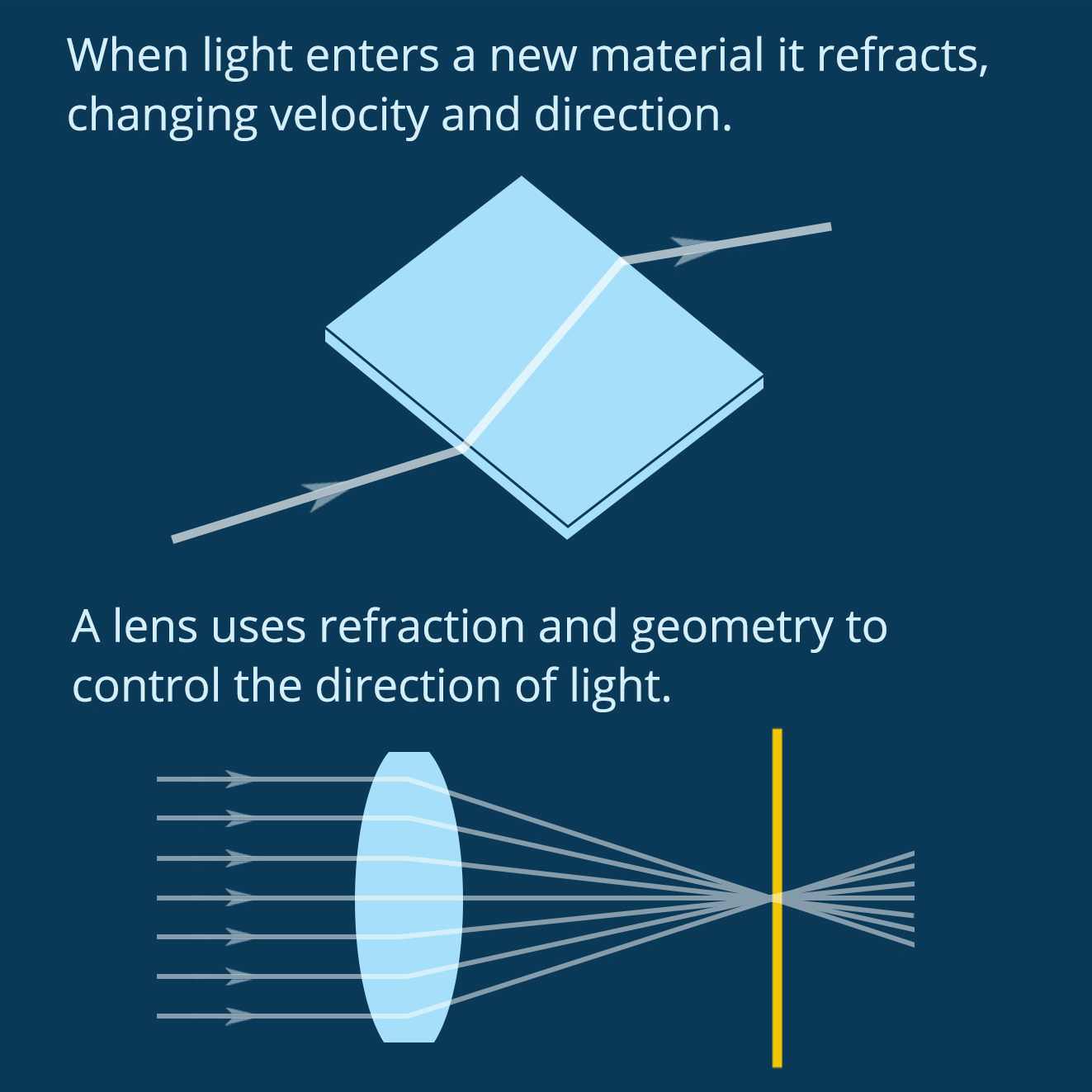
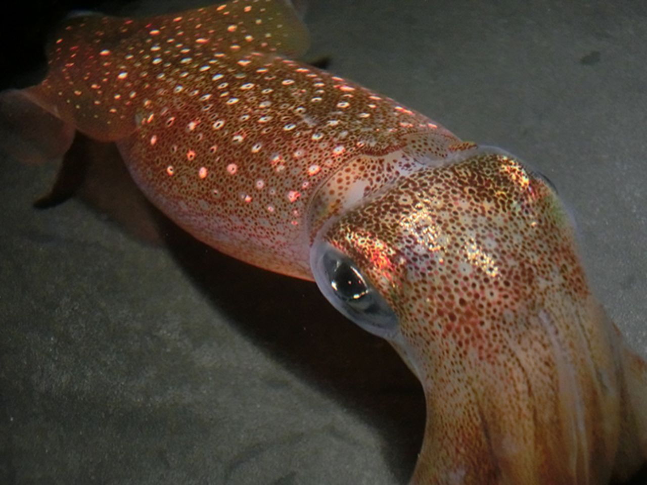
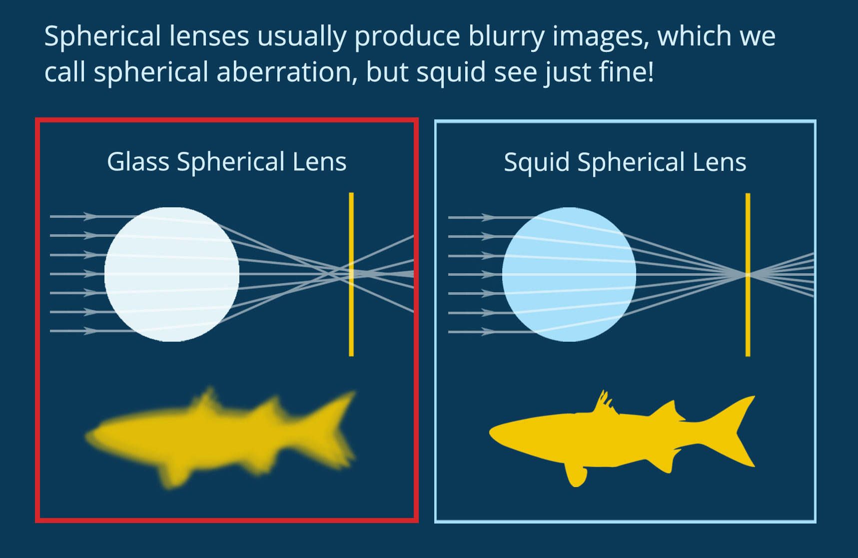
When light enters a new material it refracts, changing velocity and direction. A lens uses geometry and material properties to direct light on a specific path. You can see in Figure 1 how the shape and refractive index of a biconvex lens combine to direct light at a single spot. In biology, complex eyes like those found in most vertebrates and in squid have a lens that directs light onto the retina at the back of the eye, forming an image to be processed by the brain. Squid use spherical lenses to do this, but spherical lenses have a problem. As you can see in Figure 2, if you make a spherical lens out of one material (like glass), the light rays overlap after exiting the lens and the resulting image is blurry. This is called “spherical aberration.” Human engineers use spherical lenses a lot, and we correct for spherical aberration by combining multiple lenses. Squid, on the other hand, have evolved a lens that self-corrects for this distortion. 

Figure 2. Spherical lenses usually produce blurry images, but not in squid. (Figure by the author)

We know, in theory, how a squid might do this. In 1854, the famous physicist James Clerk Maxwell mathematically designed a spherical lens with “perfect” focus. He showed that if the density of the lens changes along the radius, forming a density gradient that he called a “perfect medium,” then the lens will produce a clear image. Today engineers can make gradient index lenses like this, but the process is difficult and energy intensive. Squid evolved to grow them easily. Could understanding how squid make these lenses help human engineers learn to do the same thing? This question inspired Dr. Jing Cai and Prof. Alison Sweeney to study the structure of the squid lens.

Figure 3. A squid lens has rings, which could combine to create a “perfect medium.” (Figure by the author, photograph from the Museum of Museum of New Zealand Te Papa Tongarewa, as reported by Nerdist)

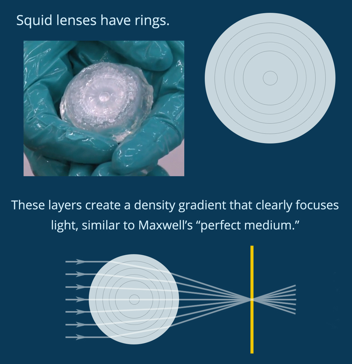
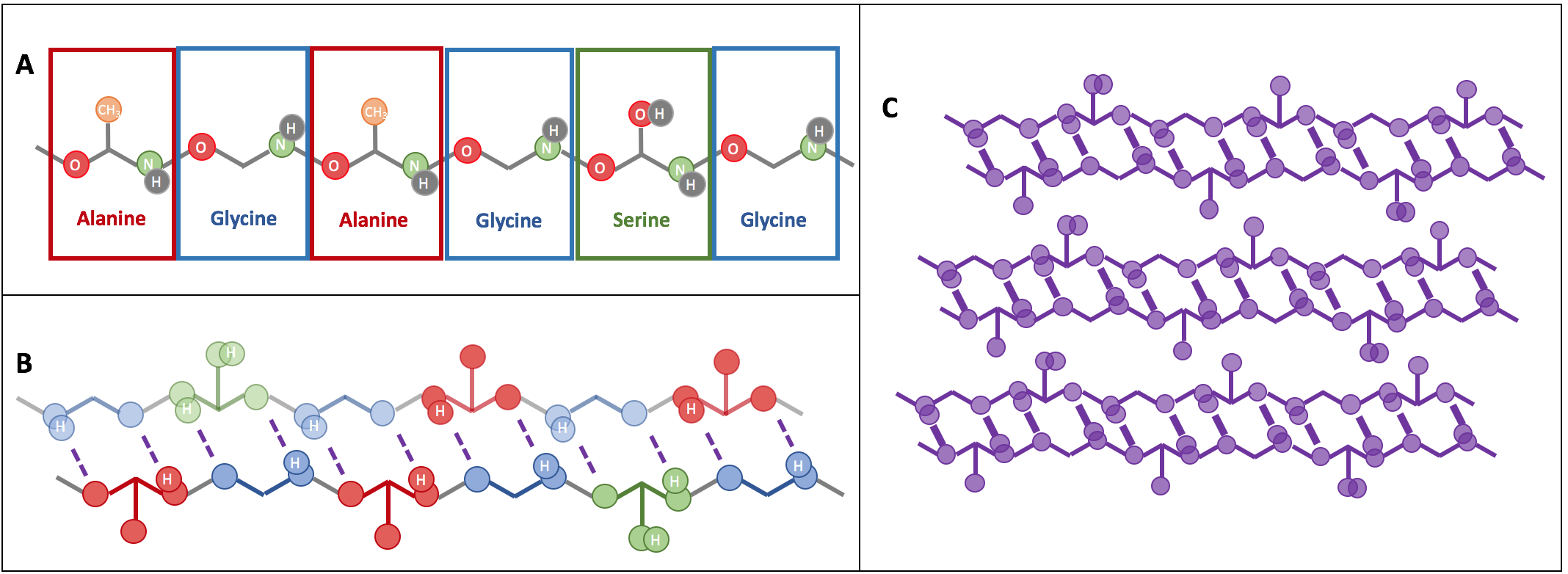
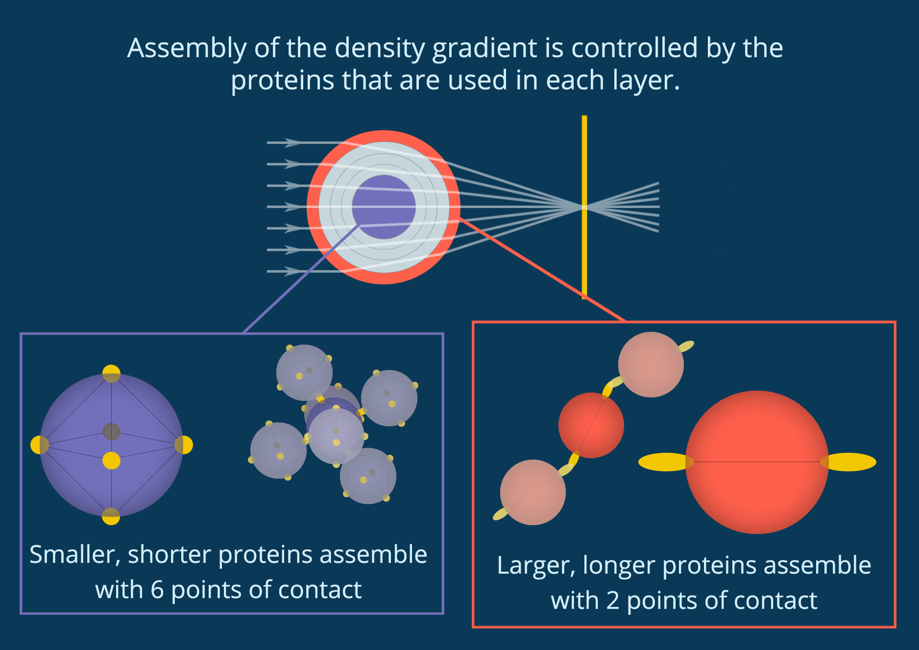
You can see in Figure 3 that when you crack open the lens in a squid eye you find rings like the inside of a tree trunk. If each of these rings has a slightly different density, they could combine to create a perfect medium. The lens of an eye is made out of proteins called crystallins, which fold into individual particles before linking together into a single material. Cai and her collaborators discovered that the lenses of the Longfin inshore squid (Doryteuthis pealeii) use 53 different crystallin proteins of different sizes. They also found that the different proteins are used in different parts of the lens, and each layer of the lens has a slightly different structure. As you can see in Figure 4, small proteins at the center of the lens are densely packed together so that each protein is connected to six other proteins. However, the larger proteins at the edge of the lens have more space between them, and each protein only touches two others. 

Figure 4. Protein particles assemble differently in different parts of the lens, creating a “perfect medium.” (Figure by the author)

This makes sense when you think about the cells that make these proteins. Cells rely on diffusion to bring building blocks to the right place for protein assembly and to send each assembled protein out to where it’s needed. When finished proteins link together to grow the lens, they disrupt this diffusion and stop protein production. By growing from dense to less dense and using so many different proteins (53 in the Longfin inshore squid), the cells are able to start and stop the growth of different layers while maintaining a single particle network. No part of the lens separates out or turns opaque, but there are still large enough regions with different densities to diffract light into alignment. 

Cai and her collaborators showed that squid lenses definitely use a density gradient similar to Maxwell’s perfect medium to correct for spherical aberration. It’s likely that this density gradient not only creates a perfect medium, but also helps control lens assembly. Now that we know how squid build a perfect spherical lens, it is easier to envision how human engineers could grow our own complex optical materials.

Anti-biofilm Material to Fight Bacterial Formation on Surfaces

Original paper: Sodium Dodecyl Sulfate (SDS)-Loaded Nanoporous Polymer as Anti-Biofilm Surface Coating Material  


Are you afraid of visiting the dentist? If so, you’re probably not the only one, but unfortunately we can’t avoid it. Yearly dental check-ups are necessary to prevent tooth and gum infections. Dentists use a disturbingly sharp, noisy tool to remove dental plaque from the tooth surface. Dental plaque is caused by bacteria, and it is an example of a biofilm, which is a community of bacterial cells that stick to surfaces. Biofilms can be found everywhere, especially on wet surfaces. Biofilms cause health problems for millions of people worldwide every year, primarily because of infections during surgery or consumption of contaminated packaged foods. To prevent these problems, some scientists are developing surface coatings that will prevent biofilm formation in the first place. In this week’s paper, we will learn about a new technique for creating a microscopic “shield” against the formation and growth of biofilms.

A biofilm is a complicated microscopic world in which multiple bacterial species can coexist. Most parts of the biofilm are covered by a protective sticky slime that is produced by the bacteria themselves. It is now known that bacteria communicate with each other within the biofilm by exchanging small molecules, proteins, genes, and even electrical signals. This intercellular communication results in expression of specific genes throughout the bacterial community in response to the environment. As a result, bacteria in biofilms are able to quickly develop resistance to antibiotics, making the treatment of biofilm infections extremely challenging. Therefore, one of the most common ways of destroying biofilms is a mechanical removal by scraping. This explains why we can’t avoid going to the dentist at least once per year; a toothbrush is not strong enough to remove the biofilm layer that we know as dental plaque.

E.coli-colony-growth.gif
Figure 1. A culture of E. coli growing. Video courtesy of en.wikipedia.org.

Unfortunately, scraping is not always possible; especially in cases where biofilms are formed at surfaces inside the body or on tiny surgical and industrial tools. Li Li and co-workers from the Technical University of Denmark, in collaboration with the Nanyang Technological University in Singapore, developed a coating material with the ultimate goal of preventing the growth of multiple bacterial species. This material has a structure with many nanoscale pores (holes) able to load and release antimicrobial compounds. For this study, it was filled with a detergent compound that is part of household cleaning products and is known to kill bacteria by dissolving the bacterial cell membrane. The researchers loaded the nanoporous polymer films with the detergent and placed the films in contact with Escherichia coli (E. coli) biofilms. Before showing what happened to the E. coli biofilms, let’s discuss a little bit more about the nanoporous polymer film.

figure 1 gyroid
Figure 2. The periodic gyroid structure of the nanoporous polymer films. Video courtesy of en.wikipedia.org.

The nanoporous film used in this study is made of a polymer that has two hydrophobic (water-repelling) chains, one of which is a silicon-based material that we use in contact lenses. The polymer chains self-organize into the beautiful gyroid structure shown in Figure 2, which is a 3D interconnected surface that repeats in three directions (is triply periodic) and contains no straight lines. The most beneficial part of this structure is that it forms small, nanoscale pores after the removal of the silicon-based chains, which provide large storage space for the detergent molecules. To stabilize the final structure used in this study, the researchers add a chemical compound to remove the silicon-based chains. At this step, the interconnected polymer chains form strong covalent bonds with each other, a process called cross-linking. Figure 3 shows the process of the nanoporous film preparation (a, b) and loading of the detergent (c, d) (to learn more about the preparation, see [1]).

figure 2 nanoporous film
Figure 3. Representation of making the nanoporous polymer film and loading it with detergent by diffusion: (a) the block copolymer re-organizes into a gyroid structure, (b) the silicon-based polymer chains are removed from the nanoporous film, (c) The detergent solution is in contact with the nanoporous film and the detergent molecules attach to the pore walls (the enlargement shows that excess free detergent molecules may form small spheres between the walls), (d) the final nanoporous film loaded with detergent (red color represents the detergent layer). (Image adapted from Li Li’s paper).

What happened to the E. coli communities after being in contact with the nanoporous film loaded with detergent? The researchers tested three samples of films differing in thickness (0.5mm, 1mm, and 1.5mm). They took microscopic images after two days and after seven days of contact with the bacteria. These specific periods were chosen because it is known that within three days almost 70% of the detergent can be released from the nanopores. To compare, they also included a nanoporous surface without detergent, which is shown in Figure 4, parts A and F. On the samples without detergent the bacteria were free to grow into large biofilms. The results in Figure 4 show the astonishing difference between the biofilms with, and without contact to the detergent after two days. Only a few small areas of live bacteria (green spots) were visible on the films with detergent, and even some dead bacteria were visible (red spots). The nanoporous surface worked! In addition, thicker nanoporous surfaces were even more effective against biofilm growth, because they have more pores loaded with detergent.

figure 4 fixed
Figure 4. Images of the 2-day (A–E) and 7-day (F–J) biofilm formation by Escherichia coli on nanoporous films with (B–E, G–J) and without (A, F) detergent. Green and red cells correspond to live and dead cells, respectively. (Image adapted from Li Li’s paper).

The tests after seven days were not as successful for the thinner films, which means most of the detergent was released from these films in less than seven days. Interestingly, the thickest nanoporous film was still effective at preventing biofilm growth after seven days. The researchers also tested the material on biofilms made by another type of bacteria, Staphylococcus epidermidis, which has a different type of cell wall. The results were not successful, and the biofilm kept growing, showing that the particular detergent is not effective in killing this type of bacteria. This shows the challenges researchers are facing, such as releasing antimicrobial compounds for longer periods of time and preventing the growth of specific bacterial species.

To conclude, this study showed that these gyroid nanoporous surfaces are effective in delivering detergent to prevent the formation and growth of E. coli biofilms. The researchers recommend further experiments with different types of detergents to target more species of bacteria. Of course, we can’t use detergents for applications in the body (detergents are highly toxic), but it is possible these nanoporous films could be used to deliver other non-toxic, antibacterial molecules. The research on the fight against biofilms keeps going! But you still have to visit your dentist every six months.


Continue reading “Anti-biofilm Material to Fight Bacterial Formation on Surfaces”

Scientists dream of micro-submarines

Original paper: Graphene-based bimporphs for micron-sized, autonomous origami machines


In the 1966 movie Fantastic Voyage, a submarine and its crew shrink to the size of a microbe in order to travel into the body of an escaped Soviet scientist and remove a blood clot in his brain. The film gave viewers a glimpse into a possible future where doctors could treat patients by going directly to the source of the problem instead of being limited by the inaccessibility of most parts of the human body. This dream of a tiny submarine that can be piloted through the human body to deliver medical care remains, even 50 years later, in the realm of science fiction. However, Miskin and coworkers at Cornell University have brought us one step closer to making this a reality with their recent development of autonomous microscale machines.

To live up to its name, an autonomous machine must have two features. First, it should be able to detect a stimulus from its environment. Then, without any help or intervention, it must respond to the stimulus with a desired response. In this scenario, the machine is not thinking or making decisions— instead, its response to the stimulus is pre-programmed. The ability to respond without supervision means that it can function in remote, inaccessible places, such as deep inside the human body.

One of the biggest challenges to miniaturizing machines is that they contain moving parts. Even fairly simple mechanisms like hinges and valves are too difficult to make on such a small scale. They would require sub-micron machining precision that is not possible using techniques available today. As a result, scientists and engineers must develop alternative mechanisms to perform the functions of these moving parts.

To address this problem, McEuen and Cohen develop a bimorph actuator— a mechanism that allows the machine to move in response to a stimulus, but does not have any complicated moving parts to fabricate [1]. Instead, the bimorph actuator is just a very thin sheet with two layers, one of graphene and the other of glass, that bends in response to changes in temperature or electrolyte concentration. The glass layer expands or contracts when exposed to the different environmental conditions [2], but the graphene does not.  The expansion or contraction of only one of the layers causes the whole sheet to bend (as shown in Movie S2) [3]. Although glass seems like a material that would break instead of bending, the actuator is only two nanometers thick so it bends easily.

nanosubmarines_fig1
Figure 1: The rigid panels on the bimorph sheet direct it to fold into the desired shape. (Adapted from Miskin et al., PNAS 2017)

To harness the motion generated by their bimorph actuator, the researchers take inspiration from an old technique: origami. Since the 17th century, origami has been used in Japan to transform flat sheets of paper into three-dimensional sculptures using only a series of folds. With paper origami, the person making the folds knows where they need to go to make the right final sculpture. However, for a micro-machine, these folding instructions must be programmed into the flat sheet during fabrication so it can fold itself. To do this, the researchers attach thick, rigid panels to certain areas of the bimorph sheet, as shown in Figure 1. The sheet is then only able to fold in the areas between the panels, so the folds are constrained by the shapes and locations of the panels. Using this technique, the researchers construct a variety of structures including a helix, a tetrahedron, a cube, and even a book with clasps, as shown in Figure 2.

nanosubmarines_fig2
Figure 2: Using bimorph actuators, the researchers make complex three-dimensional figures. On the left, the unfolded structure. Center, the folded structures, all shown with the same scale. Right, the same structure folded from paper. (Adapted from Miskin et al., PNAS 2017)

While a self-folding cube is still a long way from a submarine, this technology does open the door to the development of small machines that function on the cellular level. All of the materials used in the origami micro-machines are biocompatible, so they are non-toxic to cells yet robust enough to withstand the conditions inside the body. The closed structures could potentially be used in the body to selectively deploy a drug in response to a local environment.

With further refinement, these machines have the potential to do more complex things. They are strong enough to support electronics and still be able to fold. In fact, the faces of the folded structures are large enough to contain a microprocessor with about 30 megabits of memory or even a functional radio-frequency identification (RFID) chip. The graphene layer in the bimorph also retains its electrical properties, which may allow for the creation of a network of electrically-connected origami machines that can do more complicated tasks than one machine on its own. So, while these origami machines may be simple, they are a step toward precise sensing and manipulation of matter on the cellular scale and—maybe someday—a microscopic submarine.


[1] Bimorph, meaning “two-shape” or “two-form”, refers to the two layers of different materials. In this case, one of the materials responds to changes in the environment to produce bending. In general, either one or both materials can be active. Bimorphs are commonly used for actuation, or generating motion, as shown in this paper. They can also be used for sensing by making one of the materials is piezoelectric so it generates a voltage when it bends.

[2] The ion exchange process is well-known for being able to swell glass and is used commercially to make chemically toughened glass. In certain electrolyte or pH conditions, alkali metal or hydronium ions can diffuse into the voids in the glass and associate with dangling silicon-oxygen bonds. If the ion is larger than the pre-existing void, this causes the glass to swell. Larger ions, such as potassium, result in more swelling than smaller ions like sodium.

[3] This is the same bending mechanism by which a bimetallic strip can be used in a thermostat. The strip, which is made out of two metals that expand differently due to temperature, is made into a coil whose curvature then depends on the temperature and tells the thermostat when to adjust the temperature and in what direction.

Self-assembling silk lasers

Rings, spheres, and optical resonators self-assembled out of silk

Original paper: 3D coffee stains


When I first learned about the coffee ring effect I thought it was super cool, but it seemed like an open-and-shut case. Why do rings form where some liquids, like spilled coffee, are left to dry? Roughness on the table causes the liquid to spread imperfectly across the surface, pinning the edges of the droplet in place with a fixed diameter. Because the diameter of the droplet can’t change during evaporation, new liquid must flow from the droplet’s center to the edges. This flow also pushes dissolved coffee particles to the edges of the droplet, where they are left behind to form a ring as the water evaporates away (Figure 1). More details can be found in our previous post, here. It’s a complicated phenomenon, but after being described in 1997 it doesn’t seem like anything new would be going on here. Right? Well, as usually happens in science, classic concepts have a way of popping back up in unexpected ways. Last year It?r Bak?? Do?ru and her colleagues in Prof. Nizamo?lu’s group at Koç University, Turkey published a study using the often troublesome coffee ring effect to their advantage: making self-assembling silk lasers.

pinning
Figure 1: Pinning and the Coffee Ring Effect. A cross section of a water droplet drying on a smooth surface (A) versus a rough surface (B). On a smooth surface the droplet shrinks due to evaporation. On a rough surface the edge of the droplet is pinned and cannot shrink, forcing an internal flow to maintain the droplet’s shape.

The fundamentals here are the same as the classic coffee ring effect, but instead of coffee particles Do?ru’s droplets hold a colloidal suspension of silk fibroin proteins. In a colloidal suspension, particles (such as proteins) are mixed in another material (such as water) and neither dissolve fully into solution nor precipitate out. Smoke, milk, and jelly are all examples of colloids. Harnessing the coffee ring effect to build 2D structures out of colloidal particles has been well developed since Witten’s description of the coffee ring effect in 1997 [1], but 3D self-assembly is much less common. What makes Do?ru’s 3D structures possible is the fibroin protein.

Fibroin is the primary component of silk from the silkworm Bombyx mori. These fibers have been used by humans for thousands of years to make textiles, but recently the fibroin protein has taken on new life when extracted from silk as an aqueous, water-based, suspension and regenerated into other forms [2,3]. Fibroin proteins are long, and they easily tangle up and bond to each other to form networks of layered crystalline structures called beta-sheets (?-sheets) (Figure 2). These sheets give silk fibers and other fibroin materials strength and toughness. Furthermore, fibroin materials are biocompatible and biodegradable.

Silk Fibroin and Beta Sheets
Figure 2: Silk Fibroin And ?-sheets. Silk is made of long fibroin proteins (a) that have a repeating molecular structure. These proteins bond together into ?-sheets (b), which then stack together (c) to form materials with high strength and toughness.

To create 3D structures with the coffee ring effect, Do?ru, Nizamo?lu, and their coworkers put droplets of silk solution on superhydrophobic surfaces. Superhydrophobic surfaces strongly repel water, preventing water-based liquids from spreading flat across the surface and making the droplets stand straight up during the drying process. This makes the angle between the edge of the droplet and the surface (called the contact angle) particularly high, between 95-145 degrees throughout evaporation. The interaction between water and the superhydrophobic surface determines the shape of the final structure, with high contact angles creating more spherical droplets (Figure 3). After a solid 2D ring of fibroin forms on the bottom, the silk proteins continue to stack along the droplet’s surface, forming a stable spherical shell of ?-sheets that the remaining water can evaporate through. The researchers found that the concentration of the fibroin solution was important for controlling the final structure. If the solution is too dilute then the shell will collapse in on itself, but if the fibroin concentration is too high the initial contact angle will be lower and the final structure will also be more 2D than 3D.

Contact Angle
Figure 3: Contact Angle. Droplets of the same solution show different contact angles on different surfaces (as adapted from Do?ru’s paper). On the left is a mildly hydrophobic surface, and on the right is a superhydrophobic surface. Note how the size of the contact angle (shown in white) increases with the hydrophobicity of the surface.

To make 3D spheres, the researchers tried the pendant drop method, hanging a droplet from the tip of a needle. Similar to getting high contact angles from a droplet on a hydrophobic surface, hanging a droplet from a needle gives that droplet a small contact area, and a spherical shape (Figure 4). If the diameter of the needle is the same size or smaller than the contact area of the droplet on a superhydrophobic surface, then the shape of a droplet squeezed out of the needle should be as or more spherical than the droplets in the previous experiment. In this study, the pendant drop method ends up producing more uniform drying. These pendant-drop shells are smooth enough inside to act as optical resonators, surfaces that reflect light waves back on themselves so the waves amplify each other (the “a” in “laser,” which I always forget comes from the acronym for “light amplification by stimulated emission of radiation”).

As a proof of concept, the researchers made shells out of fibroin mixed with green fluorescent protein (GFP). Fibroin ?-sheet formation is so stable that it still happens when small amounts of other materials are present, so the optical resonator can form in the same way it did with a fibroin-only solution. In this case, because GFP has been added, when the structure is exposed to the right light source it will amplify green light emitted by the shell itself – an “all protein laser” in the making.

Benefits of the Hanging Pendant Drop
Figure 4: Benefits of the Hanging Pendant Drop. The hanging pendant drop method can produce similar spherical drops to a hydrophobic surface. It was shown that the pendant drop method produces more spherical final structures (adapted from Do?ru’s paper).

Part of what’s exciting about this publication is that the authors harness the coffee ring effect for a fun new type of small scale, self-directed 3D “printing.” They showed that the method works for other polymers as well, but I agree with their choice to highlight the silk protein fibroin. Not only is fibroin biocompatible, but it also has the potential to be more environmentally friendly to process than other polymers and is already produced in large quantities globally as part of the textile industry.


[1] Han, W. and Lin, Z. “Learning from ‘Coffee Rings’: Ordered Structures Enabled by Controlled Evaporative Self-Assembly.” Angew. Chem. Int. Ed. 51 (2012): 1534–1546.

[2] Altman, G.H. et al. “Silk-based biomaterials.” Biomaterials 24 (2003): 401–416.

[3] Koh, L.-D. et al. “Structures, mechanical properties and applications of silk fibroin materials.” Prog. Polym. Sci. 46 (2015): 86–110.

Soft nanoparticles: when polymers meet soap

Original paper: Self-Assembly of Complex Salts of Cationic Surfactants and Anionic? Neutral Block Copolymers. Dispersions with Liquid-Crystalline Internal Structure  


For more than four decades, scientists have been investigating the properties of small objects dispersed in solutions. Some of these objects – produced in laboratories – are the so called soft nanoparticles. The name soft comes from the fact that these particles are partly solid and partly liquid. One of the scientists’ aims is to design nanoparticles that will be used as carriers of medical compounds (like drugs, DNA segments, and enzymes). The nanoparticles’ role will be to protect this cargo from partial degradation through the human body until reaching the specific target cells where the nanoparticles’ structure will break up and the useful compounds will be released. This technology will allow for disease treatments using smaller amounts of drugs, which will mean fewer side effects for the patients.

The effectiveness of this treatment depends on several factors that control the nanoparticles’ properties. It turns out that one important factor is the protocol of preparation, which is the recipe used to make the nanoparticles. Today’s paper by Leticia Vitorazi, Jean-Francois Berret and Watson Loh introduces an alternative method of preparation and shows how the individual chemicals that are chosen for creating the nanoparticles can influence the nanoparticles’ properties.

The formation of the soft nanoparticles is a spontaneous process that takes place in specific mixtures of solutions because of the electrostatic attraction between two different compounds, macromolecules and surfactants. Each of these compounds usually exist as individual molecules in water (or other solvents).

Macromolecules consist of long, chain-like synthetic molecules. They can have either one long chain (called homopolymers) or two long chemically different chains connected together by covalent bonds (called diblock copolymers) (Figure 1).


figure 1.homopolymers
Figure 1. Representation of a homopolymer with a long charged chain and a diblock copolymer with a charged (A) and a non-charged (B) chain. Each sphere represents a repeated unit of the chain.


The other type of compound, surfactants, comprise short molecules with both hydrophilic (water-loving) and hydrophobic (water-hating) parts. Surfactant is what soap is made from, and its name is a shortcut to the term surface active agents. When the amount of surfactant molecules in a water solution exceeds a specific number (above the so called critical micelle concentration), the surfactants clump together and form small spheres (called micelles) with the hydrophobic parts inside the sphere to avoid contact with the water molecules, leaving the hydrophilic parts at the surface to be in contact with the water (Figure 2).


Figure 2 micelles
Figure 2. Representation of surfactant molecules with a hydrophobic tail and hydrophilic head (red spheres) surrounded by oppositely charged small ions (counterions). Above a specific concentration of surfactant in water, the surfactants molecules organize themselves in micelles with the tails inside the micelle to be protected from water and the heads in the micelle surface to be in contact with water.


Mixing macromolecule solutions with surfactants/micelle solutions causes the two compounds to come together because their opposite charges attract, and a new object is created: a nanoparticle, composed of macromolecular chains surrounded by surfactant micelles. If the macromolecule is a diblock, with a second type of non-charged chain attached chemically to the charged chain, the resulting nanoparticles usually have a specific structure with an internal core consisting of surfactant micelles and oppositely charged macromolecule chains, surrounded by an external shell. This shell consists of the macromolecules’ non-charged chains and acts as a non-stick coating that prevents the particles from clumping together (Figure 3).


Figure 3. Nanoparticles scheme
Figure 3. Schematic of soft nanoparticles with an inner core comprised of charged surfactant micelles (red), and oppositely charged polymer chains (yellow) surrounded by an external shell comprised of non-charged polymer chains (green). The red dots represent the surfactant micelles that form inside the nanoparticle core at a cubic order (enlarged scheme). Adapted from Vitorazi and colleagues.


The most popular method of creating the nanoparticles is the direct mixing method of the macromolecules and surfactants. The two compounds are dissolved separately in water (or an appropriate solvent), and then they are mixed together to reach specific amounts of the opposite charges in the solution. The mixtures consist of charged macromolecules and oppositely charged surfactants surrounded by small ions. The resulting nanoparticles are small: between 30 and 50 nm. (For comparison, HIV virus is about 120 nm large and bacteria are about 1000 nm.)

Vitorazi and co-workers used a different method named “complex salts.” The recipe of making the complex salts consists of mixing the macromolecule solutions with an increasing amount of the surfactant solutions until all acidic groups of the macromolecules bind to the hydroxide groups of the surfactants. The next step of the process is the removal of the solvent to create a single-component powder, which is freed from the small ions that surrounded the individual compounds in the solution (Figure 4A). Finally, the powder is dissolved in water at different concentrations and the electrostatic attraction between the surfactant micelles and oppositely charged macromolecules results in the formation of the nanoparticles.


Figure 4. the complex salts vs direct mixing
Figure 4. Representation of a diblock copolymer with charged (yellow parts) and non-charged (green parts) chains surrounded by surfactant micelles (red parts) in the absence of small ions (A) and in the presence of small ions (B).


The researchers used the complex salts method for different macromolecular chain lengths to explore the effect of the macromolecules’ chain lengths on the nanoparticles’ size and core structures, and they compared the results with the direct mixing method that was used in previous years for similar mixtures. They found that if the two sub-chains of the diblock copolymer (A and B in Figure 1) have roughly equal lengths then the nanoparticles were larger, compared to those made through the direct mixing method. As shown in Figure 3, these particles consist of a core with a cubic ordered structure where the surfactant micelles are positioned at specific places in the core. They also found that for unequal sub-chain lengths, the core was disordered and the nanoparticles were smaller. This is an important finding because the ordered structure and the larger size of the nanoparticles can incorporate larger amounts and different types of drugs in the core.

They also studied the effect of the addition of salt or macromolecular solutions in the nanoparticle solutions. The amount of salt strongly affects the nanoparticles’ properties because the added ions in the nanoparticles’ solution weaken the attraction between the macromolecules and the oppositely charged surfactant micelles. Therefore, it is a useful factor to be considered for the biomedical applications in the salty environment of the cell system in our body. Vitorazi and co-workers found that after the addition of salt or macromolecule solutions in the nanoparticles’ solution, the already formed nanoparticles lost their internal cubic structure (meaning that the micelles were now randomly oriented in the core) but the stability of the system was preserved.

To conclude, the method of preparing soft nanoparticles plays an important role in determining their properties and therefore affects their performance as drug carriers. Compared to direct mixing methods, the complex salts formed stable and 3 times larger nanoparticles with a core of cubic internal order. In a charged environment (created by addition of small or macromolecular ions) the final structure lost its cubic structure, but the nanoparticles were still stable, making them important candidates for the future of drug delivery technology.