Bioinspired e-skins: camouflaging with the flip of a switch

Original paper: Bioinspired MXene-Based User Interactive Electronic Skin for Digital and Visual Dual Channel Sensing   

Content review: Heather Hamilton
Style review: Arthur Michaut


Human skin has many functions beyond ensuring that all of our insides stay, well, inside. Skin also acts as a giant sensor that feels sensations like pressure, temperature, or vibration, and converts them into electrical signals to be processed by the brain. In the animal kingdom, some species like chameleons can even use their skin to selectively blend into their environments. Scientists have set out to create electronic skins, or e-skins, that can mimic or even outperform the typical functions of the human skin by taking on color-changing abilities like chameleon skin. Polymer materials are excellent for mimicking skin due to their soft and elastic nature that allows them to bend and stretch without tearing. However, most polymers are not conductive, a necessary property for transporting electrical signals. To create e-skin, conductive materials are often integrated into polymer materials to add electronic properties to a flexible skin-like matrix. By also integrating a color-changing pigment, researchers from Tongji University created a unique e-skin that could digitally via measurement of current flow and visibly detect mechanical movements and change color for effective camouflaging. 

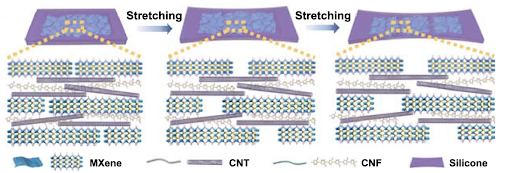
Conductive, metal-based nanosheets, called MXenes, were used because of their hydrophilic quality, which means they like water similar to our skin cells. When encapsulated in the polymer matrix, the nanosheets are connected and can conduct electrical current. However, when the e-skin stretches, the nanosheets become disconnected and stop conducting the current. To remedy this, smaller conductive materials called carbon nanotubes were mixed with cellulose nanofibers and added to the e-skin. The cellulose nanofibers (CNF) help disperse the carbon nanotubes (CNT) similar to how coating fruit with flour before adding to cake batter helps the fruit disperse in the cake during baking rather than sinking to the bottom. The carbon nanotubes act as a bridge between nanosheets in stretched states as seen in Figure 1.

Figure 1. Schematic of conductive e-skin components in response to stretching.

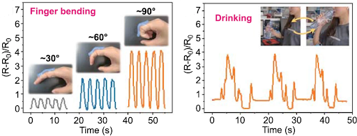
The researchers measured the resistance to electrical current flow, or resistivity, to determine the e-skin electrical response to stretching. The difference in electrical current between an unstretched and stretched e-skin can be used to detect large movements, like a finger bending at different angles, and subtle movements, like throat movements while swallowing. As shown in Figure 2, the e-skin exhibits different relative resistance to the flow of the current at different finger bending angles. 

Figure 2. Signal responses in the form of relative electrical resistance, (R-R0)/R0, showing mechanical movements can be detected by e-skin.

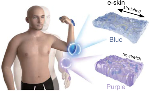
From their experiments, the researchers realized that the e-skin may have Joule heating capabilities meaning the e-skin would heat it up when current is flowing through it and quickly cool down as soon as the current is turned off. Inspired by color-changing abilities found in some animal skins, the researchers also mixed thermochromic pigment into the polymer matrix before casting the e-skin. These pigments change color as they are heated above 31°C. (Figure 3a).

Figure 3. E-skin containing thermochromic pigment in response to (a) Joule heating and (b) stretching.

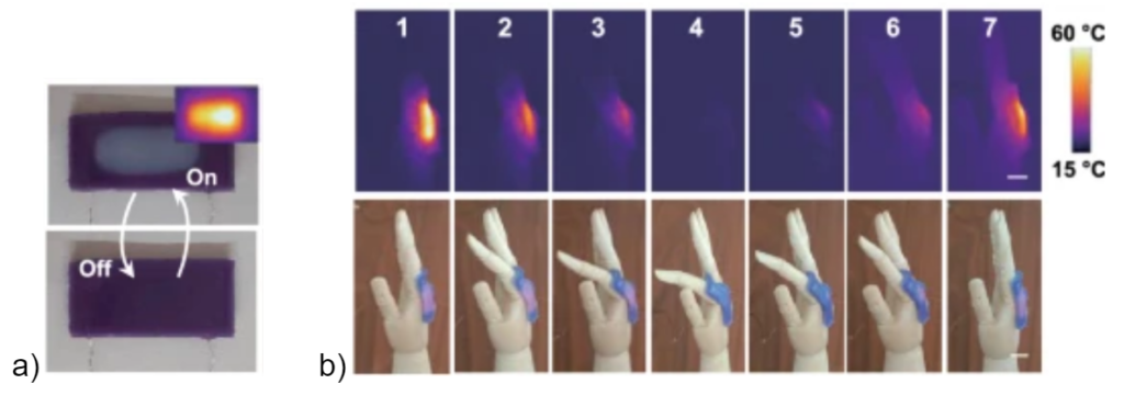
As soon as the current is turned off, the e-skin begins to cool, returning back to its initial color. Since the current flow can be altered by stretching the e-skin, the temperature and subsequent color can also be changed by stretching the skin. When current is flowing through the unstretched skin, the e-skin temperature increases, causing it to turn white. When stretched, an increase in electrical resistance causes the e-skin to return to its initial color. Instead of waiting for an electrical current reading, strain and temperature are visibly detected by the color of the e-skin. This strain color-changing ability was used to detect finger movements when the e-skin was applied on a hand (Figure 3b). To demonstrate the camouflage phenomenon similar to a chameleon changing its colors to mimic the environment, an e-skin was attached to a green plant (Video).

Video. E-skin changing color to match the color of the green plant.

The e-skin changes color from a dark green to a light green similar to the plant in about 90 seconds simply by turning on a current. This e-skin camouflaging ability goes well beyond the natural abilities of human skin and would be useful for military applications for quick camouflaging. The creation of this multifunctional e-skin is an exciting innovation for the wearable technology field and brings us one step closer to becoming chameleons.

The Future of Shape-Memory Polymers: Just Add Water and Glycerol

Original paper: Magnetically Addressable Shape-Memory and Stiffening in a Composite Elastomer


Video 1. Heat-shrink tubing demonstration.

Have you ever used heat-shrink tubing at home to seal an exposed wire? As shown in Video 1, you would place the tubing around your wire, apply heat, and voilà! The tubing shrinks and tightly wraps itself onto the exposed wire, and you don’t have to worry about an electric shock anymore. This type of material that changes its shape upon increased temperature is called a shape-memory polymer. Since its commercial development in 1962, scientists have found this type of material so useful that its popularity rose, especially in the biomedical and aerospace fields. However, it comes with a few drawbacks: applying the desired temperature uniformly can be tricky and the shape change induced by the heat can be quite slow. In addition, changing the temperature isn’t ideal for biological applications where the environment surrounding the material is sensitive to heat, such as in tissues and living cells. In today’s post, I’ll introduce you to a different type of shape-memory material that “remembers” its temporary shape when subjected to a magnetic field, instead of heat.

Video 2. Demonstration of the material developed by the authors.

Paolo Testa and coworkers from the Paul Scherrer Institute and ETH Zurich developed a shape-memory polymer composite that can preserve a new shape when a magnetic field is present. As you can see in Video 2, the initially flexible material sitting on the center stage is manually twisted with a tweezer and held by force. When the magnetic ring is raised around the stage and held up so that the material can “feel” the magnetic field, the material “freezes” in its twisted shape, even when the tweezer is removed. After a certain time, the magnetic ring is removed and the material regains its original shape within one second, dramatically faster than the time it took for a heat-shrink tubing in Video 1 to change its shape. The material—at least a part of it—that looks like black rubber is a flexible polymer that is the main ingredient of the Silly Putty, called poly(dimethylsiloxane) (PDMS). However, PDMS is normally transparent in color, and PDMS itself isn’t a shape-memory polymer. So what makes it black and capable of holding the twist when the magnetic field is present?

The answer is in iron particles. However, iron particles alone cannot perform well enough. Shape-memory materials that were previously developed had iron particles directly embedded in the polymer but didn’t have such a high sensitivity to magnetic fields. What makes the material in this paper so unique is the liquid surrounding the particles. The iron particles are dispersed in a water and glycerol mixture, making the fluid six times more viscous and stiff when subjected to the magnetic field. This type of fluid, called a magneto-rheological fluid, is then injected into the PDMS polymer, making the material sensitive to the presence of a magnetic field.

3D visualization of the material and 2D slices of that visualization with and without magnetic field
Figure 1. (a) 3D visualization of the PDMS injected with magneto-rheological fluid. (b)(c) 2D slices of a droplet surrounded by PDMS with the magnetic field (b) off and (c) on. (Adapted from the original paper.)

Figure 1 shows the 3D structure of the developed material, as well as a 2D schematic of a magneto-rheological fluid droplet with and without the magnetic field. When the fluid is injected into the polymer, the polymer encases the fluid and a composite is formed, which is shown in Figure 1a. In the absence of the magnetic field, shown in Figure 1b, the iron particles are dispersed and mobile inside the fluid droplet. However, when the magnetic field is turned on, shown in Figure 1c, the particles reorganize and align along the direction of the magnetic field, and, in turn, the droplet stiffens. The alignment of the iron particles and the resulting stiffening of the fluid, induced by the presence of a magnetic field, are the reasons why the polymer composite can hold its new shape (as in Video 2). When the magnetic field is removed, the particles regain their mobility and the fluid droplets soften, which lets the polymer return to its original shape.

On the left, a graph of stiffness as a function of the fluid volume fraction. In the middle, a graph of connectivity as a function of the fluid volume fraction. On the right, two 3D construction of material's internal structure, one representing 10% fluid and the other representing 40% fluid.
Figure 2. (a) The volume fraction (ratio) of magneto-rheological fluid over PDMS (?) versus the material stiffness. The blue and red lines indicate the magnetic field being on and off, respectively. (b) The volume fraction (ratio) of magneto-rheological fluid over PDMS (?) versus the connectivity between the fluid droplets. (c) 3D reconstructions of the material’s internal structure when the fluid occupies 10% (top) and 40% (bottom) of the total volume. PDMS and the fluid are represented in grey and red, respectively. (Adapted from the original paper.)

By controlling the ratio between the magneto-rheological fluid and PDMS (?), the authors tried to understand the relationship between the stiffness and structure of the material and the said ratio. When the percentage of the fluid increases, as shown in Figure 2a, the overall stiffness of the material measured in the presence of the magnetic field increases as well. This increase is enhanced when fluid occupies more than 20% in volume, as highlighted in yellow in Figure 2a. Compared to a factor of two increase going from 10% to 20%, the stiffness increases by a factor of 13 going from 10% to 30%.

By measuring the connectivity [1] between the fluid droplets using an X-ray scan [2], the authors discovered that the stiffness increase is related to the network connection between the fluid droplets when the magnetic field is applied (Figure 2b). This network connection is visualized in Figure 2c; the fluid, shown in red, is isolated when the fluid composes only 10%. However, when increased to 40%, the dispersed fluid pockets become connected to one another, enhancing the stiffness of the overall material under the magnetic field.

As mentioned above, the addition of the fluid was the key to the material’s drastic functional improvement. Since the fluid provides freedom for the iron particles to move around and align under the magnetic field, the stiffening process becomes more dramatic compared to having the particles alone inside the polymer. Also, the fluid acts as a buffer, lessening any damages caused to the polymer by the motion of the hard particles. The authors hope that their research will open up an even wider range of applications using this shape-memory polymer, such as a magnetic-controlled micromachine that can deliver drugs to targeted areas inside our body. Unlike the temperature-controlled shape-memory material introduced in the beginning, the stiffening of this new magnetically controlled shape-memory material is reversible. This might offer a greater potential for new applications which might require several cycles of deformation. Maybe in the near future, we’ll use items made out of magnetically controlled shape-memory polymers in our daily lives.


[1] The connectivity is defined as the volume of the biggest droplet over the total volume of the magneto-rheological fluid in the polymer composite.^

[2] The authors used X-ray tomography, a method where a 3D image is constructed using 2D X-ray images.^