Scientists dream of micro-submarines

Original paper: Graphene-based bimporphs for micron-sized, autonomous origami machines


In the 1966 movie Fantastic Voyage, a submarine and its crew shrink to the size of a microbe in order to travel into the body of an escaped Soviet scientist and remove a blood clot in his brain. The film gave viewers a glimpse into a possible future where doctors could treat patients by going directly to the source of the problem instead of being limited by the inaccessibility of most parts of the human body. This dream of a tiny submarine that can be piloted through the human body to deliver medical care remains, even 50 years later, in the realm of science fiction. However, Miskin and coworkers at Cornell University have brought us one step closer to making this a reality with their recent development of autonomous microscale machines.

To live up to its name, an autonomous machine must have two features. First, it should be able to detect a stimulus from its environment. Then, without any help or intervention, it must respond to the stimulus with a desired response. In this scenario, the machine is not thinking or making decisions— instead, its response to the stimulus is pre-programmed. The ability to respond without supervision means that it can function in remote, inaccessible places, such as deep inside the human body.

One of the biggest challenges to miniaturizing machines is that they contain moving parts. Even fairly simple mechanisms like hinges and valves are too difficult to make on such a small scale. They would require sub-micron machining precision that is not possible using techniques available today. As a result, scientists and engineers must develop alternative mechanisms to perform the functions of these moving parts.

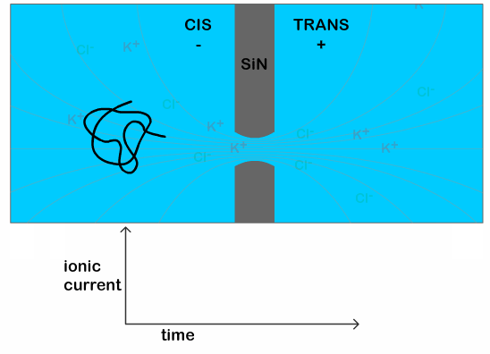
To address this problem, McEuen and Cohen develop a bimorph actuator— a mechanism that allows the machine to move in response to a stimulus, but does not have any complicated moving parts to fabricate [1]. Instead, the bimorph actuator is just a very thin sheet with two layers, one of graphene and the other of glass, that bends in response to changes in temperature or electrolyte concentration. The glass layer expands or contracts when exposed to the different environmental conditions [2], but the graphene does not.  The expansion or contraction of only one of the layers causes the whole sheet to bend (as shown in Movie S2) [3]. Although glass seems like a material that would break instead of bending, the actuator is only two nanometers thick so it bends easily.

nanosubmarines_fig1
Figure 1: The rigid panels on the bimorph sheet direct it to fold into the desired shape. (Adapted from Miskin et al., PNAS 2017)

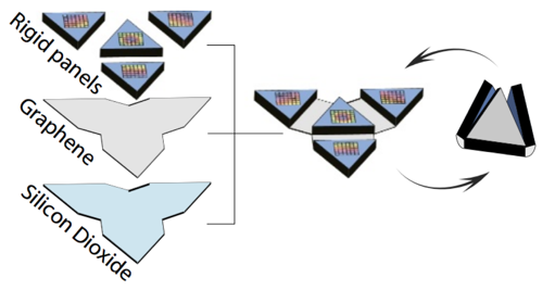
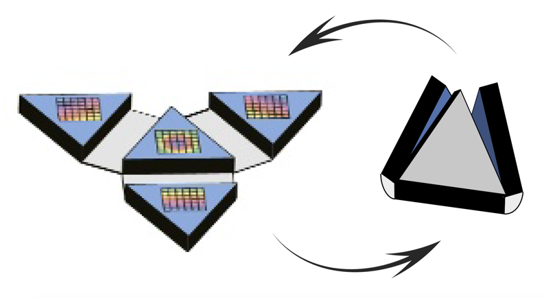
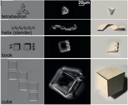
To harness the motion generated by their bimorph actuator, the researchers take inspiration from an old technique: origami. Since the 17th century, origami has been used in Japan to transform flat sheets of paper into three-dimensional sculptures using only a series of folds. With paper origami, the person making the folds knows where they need to go to make the right final sculpture. However, for a micro-machine, these folding instructions must be programmed into the flat sheet during fabrication so it can fold itself. To do this, the researchers attach thick, rigid panels to certain areas of the bimorph sheet, as shown in Figure 1. The sheet is then only able to fold in the areas between the panels, so the folds are constrained by the shapes and locations of the panels. Using this technique, the researchers construct a variety of structures including a helix, a tetrahedron, a cube, and even a book with clasps, as shown in Figure 2.

nanosubmarines_fig2
Figure 2: Using bimorph actuators, the researchers make complex three-dimensional figures. On the left, the unfolded structure. Center, the folded structures, all shown with the same scale. Right, the same structure folded from paper. (Adapted from Miskin et al., PNAS 2017)

While a self-folding cube is still a long way from a submarine, this technology does open the door to the development of small machines that function on the cellular level. All of the materials used in the origami micro-machines are biocompatible, so they are non-toxic to cells yet robust enough to withstand the conditions inside the body. The closed structures could potentially be used in the body to selectively deploy a drug in response to a local environment.

With further refinement, these machines have the potential to do more complex things. They are strong enough to support electronics and still be able to fold. In fact, the faces of the folded structures are large enough to contain a microprocessor with about 30 megabits of memory or even a functional radio-frequency identification (RFID) chip. The graphene layer in the bimorph also retains its electrical properties, which may allow for the creation of a network of electrically-connected origami machines that can do more complicated tasks than one machine on its own. So, while these origami machines may be simple, they are a step toward precise sensing and manipulation of matter on the cellular scale and—maybe someday—a microscopic submarine.


[1] Bimorph, meaning “two-shape” or “two-form”, refers to the two layers of different materials. In this case, one of the materials responds to changes in the environment to produce bending. In general, either one or both materials can be active. Bimorphs are commonly used for actuation, or generating motion, as shown in this paper. They can also be used for sensing by making one of the materials is piezoelectric so it generates a voltage when it bends.

[2] The ion exchange process is well-known for being able to swell glass and is used commercially to make chemically toughened glass. In certain electrolyte or pH conditions, alkali metal or hydronium ions can diffuse into the voids in the glass and associate with dangling silicon-oxygen bonds. If the ion is larger than the pre-existing void, this causes the glass to swell. Larger ions, such as potassium, result in more swelling than smaller ions like sodium.

[3] This is the same bending mechanism by which a bimetallic strip can be used in a thermostat. The strip, which is made out of two metals that expand differently due to temperature, is made into a coil whose curvature then depends on the temperature and tells the thermostat when to adjust the temperature and in what direction.

Microcannons firing Nanobullets

Original Paper: Acoustic Microcannons: Toward Advanced Microballistics


Sometimes I read papers that enhance my understanding of how the universe works, and sometimes I read papers about fundamental research leading to promising new technologies. Occasionally though, I read a paper that is just inherently cool. The paper by Fernando Soto, Aida Martin, and friends in ACS Nano, titled “Acoustic Microcannons: Toward Advanced Microballistics” is such a paper.

The grand scheme of this research is developing a tool that can selectively shoot drugs into cells at a microscopic level. This is hard because everything happens really slowly at the microscopic scale in a liquid, in ways that meter-sized beings who live in air would not necessarily expect. For example, it is impossible for small organisms to move through a fluid using a repetitive motion that looks the same in reverse. The way we move our feet back and forth to walk would not work for a tiny aquatic human, because the forward motion in the first phase of movement would be nullified by backwards motion in the second phase. This is why bacteria use things like rotating flagella to move*.

Digressions aside, if you tried to shoot a tiny bullet through a cell wall, it would quickly halt and diffuse away before even hitting the cell wall. Soto, Martin, and collaborators wanted to beat this. Perhaps inspired by the likely unrelated Rodrigo Ruiz Soto, a Costa Rican competitive pistol shooter in the 1968 Olympics, Soto sought to develop a cannon that would change the game in the microscopic world in the same way that gunpowder technology changed things in  the macroscopic world.

The researchers developed a “microcannon,” starting with a thin membrane of polycarbonate plastic studded with small pores, which is a thing you can buy and don’t have to make. The pores would eventually serve as the molds for the barrels of the cannons. They deposited graphene oxide onto the inside of the pores using electrochemistry, and then sputtered gold onto the inside of that graphene layer.  While they were still in the plastic membrane, the cannon pores were filled with a gel (literally gelatin from the supermarket) loaded with micron-sized plastic beads to act as bullets, and the “gunpowder,” which I’ll describe after the next image. The polycarbonate is then washed away with acid, leaving free-floating carbon and gold cannon barrels a few microns in size.

cannon
Figure 1: The microcannons, loaded with nanobullets before and after firing. Adapted from Soto and collaborators

While it is generally difficult to make small things move quickly in a fluid, bubbles are somewhat of an exception to this rule. Their collapse can lead to rapid motion on tiny scales. Taking advantage of this, the authors used perfluorocarbon (molecules with the same structure as hydrocarbons but with fluorine connected to carbon atoms instead of hydrogen) droplets as a propellant, which they turned into bubbles with an ultrasound-induced phase transition (essentially blasting them with soundwaves until they vaporized). When they initiated the collapse of the bubbles, they emitted a pressure wave which drove the nanobullets out of the barrel towards their target**.

cannon2
Figure 2: Composition and operation of the microcannons.

The authors performed two tests to characterize how powerful these things were. First, they embedded the cannons in an agar gel (an algae-based substance that Japanese desserts are made of) and loaded them with fluorescent beads. They looked at where the beads were before firing the ultrasound trigger at the cannon, and after. They observed that the beads had penetrated an average of 17 microns through the gel. However, this is about the thickness of a human cell layer, so this could be used, for example, to shoot a small amount of medication through the layer of cells on the wall of a blood vessel. In some more direct studies of the damage caused by collapsing bubbles (which is a common mechanism of damage to ship propellers), the jets that formed when bubbles collapse were shown with high-speed photography to penetrate about a millimeter into a gel. However, these bubbles were 1000 times bigger than those formed in the microcannons, and it’s not out of the question to assume that the penetration depth scales with bubble size.

jjrqrsy
Figure 3: High-speed photography of a millimeter-sized bubble collapsing near a gel wall and shooting a jet into the gel. The mechanism of nanobullet-firing and penetration is a smaller version of this. From Brujan, Emil-Alexandru, et al. “Dynamics of laser-induced cavitation bubbles near an elastic boundary.” Journal of Fluid Mechanics 433 (2001): 251-281.

The bullets were too fast to record with a microscope camera, so their second test involved recording the motion of the cannon after it fired the bullets. Naively, one would expect to be able to calculate the bullet speed with conservation of momentum from knowing the cannon’s speed, but momentum isn’t conserved in a noisy viscous environment (which brings us back to why it’s so hard for microorganisms to move around). They modeled the fluid dynamical forces acting on the system, measured that the terminal speed of the cannon was about 2 meters per second, and concluded that the initial speed of the bullets is 42 meters per second or 150 kilometers per hour (see appendix). Pretty fast, especially for something so small in a draggy environment.

After finding this paper I emailed the first author, Fernando Soto, saying that I enjoyed his paper, and he responded by saying that he was glad that other people liked his “very sci-fi nanodream.” I don’t know if this technology will succeed in the authors’ goal of localized drug delivery to cells, but I think it’s awesome that they made a functioning microscale cannon.

cannon3
Oh the humanity.

*I recommend reading Life at Low Reynolds Number if this interests you.

**Or just in whatever direction it was pointing, I guess.


Appendix: Velocity calculation

The researchers wanted to figure out how fast the bullets were moving based on their measurement of how fast the cannons were moving. Normally you could just use conservation of momentum, but because of the surrounding fluid, momentum is not necessarily conserved (unless you know the momentum of the fluid as well).

However, we understand how velocity decreases in a fluid based on drag: if the velocity is low, the drag force arises from separating the water molecules from each other, and the force is linear with velocity. If the velocity is high, the force arises mainly from accelerating the water to the speed of object, and the force is quadratic with velocity. To figure out which rule applies you can calculate what’s called the Reynold’s number, Re, which is the ratio of inertial to viscous forces in a fluid. If Re is in the thousands or higher,the flow is turbulent. f it’s below 100, the flow is smooth, or laminar. Specifically, the Reynold’s number is calculated as:

Re=\frac{\rho L v}{\mu}

where \rho is the density of the fluid, L is the length of the object in the flow, v is its velocity, and \mu is the viscosity. The microcannon was seen moving at about a micron per second, it was about 15 microns long, and the high speed photograph was done in water (density of 1 kg/L, viscosity of about 0.001 pascal seconds). This means the Reynold’s number was about 13, in the laminar regime, and that drag is due to viscosity and linear.

The equation of motion for a slowing object undergoing viscous drag with an initial velocity is

v(t)=v(0)e^{-kt/m}

where m is the mass of the cannon (known from stoichiometry) and k is the drag coefficient which depends on the viscosity as well as the geometry of the object experiencing drag. Because they know v(t) (as determined from high speed videography), t (the time since detonation), k, and m, they can find v(0).

Then it is assumed that momentum is conserved during the detonation, so the nanobullets with known mass can have their velocity calculated from

v_{c}m{c}=v_{b}m_{b}

where the indices c and b refer to cannon and bullet. The velocity was calculated to be 42 m/s. Pretty fast.


Knotty DNA

Original paper: Direct observation of DNA knots using a solid-state nanopore


Try taking out your earphones from your pocket and, in all probability, you’ll find knots and entanglements between the ends. As it turns out, this knotting effect is not limited to macroscopic objects, but occurs on the nanoscale as well. A DNA molecule that carries the genetic information of a living organism is actually a long string-like polymer, so you can imagine that it would also get tangled up just like the cords of your earphones. In fact, scientists know that DNA does form knots when it is in the nucleus of a cell, and these knots need to be removed by specialized bio-molecules, called enzymes, so that a cell can ‘read’ the genetic information encoded in the DNA. [1] In today’s paper, Calin Plesa and his colleagues at TU Delft are able to observe and measure these knots in DNA strands. In the process, they also observe interesting knotting behaviour which has not been observed before.

Knots on DNA

DNA translocation through a solid-state nanopore
Figure 1: This animation shows the DNA moving  through the nanopore. The associated dip in current is mapped onto the graph below. (Animation created by Calin Plesa, available under CC BY-SA license)

The researchers use a nanopore sensor to infer the structural properties of a DNA molecule. The sensor is made up of two reservoirs filled with electrolyte (a solution which separates into cations and anions, which can be used to conduct electricity, e.g. a salt solution), and they are separated by a membrane, or thin sheet, with a tiny hole in it. An electric field applied across the membrane generates an ionic current in the electrolyte and also pulls a negatively charged DNA strand through the tiny opening. The passage of a DNA strand through the nanopore causes a dip in the ionic current that is proportional to the volume of ions displaced—in other words, it’s proportional to the size of the molecule (a typical scenario is shown in Figure 1). Therefore, a knot in the DNA can generate a bigger drop in the current than an untangled strand. From this difference it is possible to infer the characteristics of the knot itself, since a bigger drop indicates a bigger knot.

The typical time for a DNA to pass through the pore is in the order of a few milliseconds, when the DNA is in a solution of potassium chloride (which is the typical salt solution used to carry out nanopore experiments). This makes it difficult technically, to see any features present on the DNA. Previous work has shown that it is possible to slow down the DNA passage by at least 10 times by using lithium chloride as their salt solution. [3] This increase in the translocation time (time it takes for the DNA to pass through the pore) is necessary to clearly see the additional dip in the current as the knot traverses the pore, as illustrated in Figure 2.

Translocation of a DNA molecule containing a trefoil knot through a solid-state nanopore
Figure 2: This animation shows a DNA with a knot moving through the pore. An additional dip in the current can be seen in the current trace as the knot (purple line) passes through the pore. (Animation created by Calin Plesa, available under CC BY-SA license)

The dip in the current signal caused by the knot passing through the pore can then be used to infer characteristics about the knot. In particular, it can be used to calculate the size of the knot, which has not been experimentally determined before. This has both physical and biological significance. Physically, it helps us understand the types of knots being formed on polymers as it can tell us whether the knot is loose or tightly formed. Biologically, it can help us understand how naturally occurring enzymes are able to disentangle knots in DNA strands, a function which is still poorly understood. The size of the knot is estimated by using

d= v t

where d is the length of the knot along the DNA strand, v is the average speed of the DNA translocation, and t is the time the knot takes to traverse the pore. Using this technique, the researchers estimate that the majority of the knots are less than 100 nm long. Previous research has shown that the DNA strand is rigid over lengths shorter than 50 nm, so considering this, the estimated knot size suggests that the knot is very tight. [2] However, this result needs further analysis, as the process of pulling the DNA through the nanopore might cause the knot to tighten, so this might not be the knot’s size in its natural state.

Slipping and sliding knots

When considering a linear (think: a thread with loose ends) DNA molecule, there is a possibility of the knot ‘slipping’ off the end of the strand before it gets pulled into the nanopore. For the knot to traverse the pore, it needs to be pulled fast enough to get squeezed to the size of the pore. If this process doesn’t happen fast enough the knot ‘halts’ at the pore entrance while the unknotted region translocates through. This allows the knot to disentangle, in case of a linear DNA molecule.

To determine if this slipping process occurs in knotted DNA strands, the researchers repeat their experiment using a circular (think: a thread joined end-to-end) DNA molecule. By using a closed loop they avoid possibility of the knot disentangling, but the knot can still slip towards the trailing end of the DNA during the translocation. The position of the knot is determined by the position of the dip in the current signal (purple line in Figure 2). They measure the probability of finding the knot at each position along the strand using two voltages, 100 mV and 200 mV. As shown in Figure 3, the knots show a preference for sliding toward the trailing end of the molecule at higher voltages, indicating that pulling too hard on the leading end of the DNA strand can indeed cause knots to slip along the strand instead of being pulled through the pore. The researchers also observe a 55% higher knotting occurrence in the circular molecules compared to linear ones. This suggests that knots may have slipped off the end of the linear molecules, thereby not detecting them at all.

Figure 4
Figure 3: The graph shows the probability of detecting the position of the knots along the length of DNA. At 200mV, the knots are observed to be at the trailing end of the DNA motion indicating the slipping phenomenon (adapted from Plesa et al.)

The researchers in this study have shown that naturally induced knots occur in DNA strands and they measured the sizes of those knots, which were previously unknown. This measurement showed that the knots detected are actually quite tight, which was not expected, although this result still needs to be investigated further. Additionally, these knots were seen to slide along the DNA molecules as they traversed the nanopore due to the strong pull at the end of the DNA strand. This was seen clearly by repeating the knotting experiments using circular DNA where there were no ends for the knots to slide off.

This new information about the structure of knots in DNA strands will help inform future studies of the complex topological structures formed in biomolecules such as DNA and proteins. It will also contribute to understanding the effects of topological features on the biological functions of these long, string-like biomolecules. In effect, it can help us explain the consequence of knotted DNA on the cell’s function as well as how the cell is equipped to handle these defects.

[1] http://www.tiem.utk.edu/~gross/bioed/webmodules/DNAknot.html

[2] Baumann, Christoph G., et al. “Ionic effects on the elasticity of single DNA molecules.” Proceedings of the National Academy of Sciences 94.12 (1997): 6185-6190.

[3] Kowalczyk, Stefan W., et al. “Slowing down DNA translocation through a nanopore in lithium chloride.” Nano letters 12.2 (2012): 1038-1044.