Soft Matters in Viral Pandemics

Original paper: Soft Matter Science and the COVID-19 Pandemic (arXiv here)


Quintessential soft matter problems, such as the behavior of droplets in ink-jet printing, involve complex interactions between forces and materials. In today’s article, Prof. Wilson Poon points out that coronaviruses are also quintessential soft matter objects, and highlights a range of areas where soft matter science may help better understand, and combat viral pandemics.

To a physicist, a virus is merely an inert particle that drifts around in water (it is a colloidal particle, in the jargon of soft matter physics, see Figure 1), where a single coronavirus particle is roughly spherical and around one tenth of one thousandth of one millimeter in width ($latex 0.1 \mu m$). These inert particles, however, are also covered in tiny specially shaped keys (proteins), and when these keys meet the correctly shaped lock (a receptor at the surface of a cell membrane), they activate. The previously inert particle now instructs the invaded cell’s machinery to make more viruses (imagine a Ford car whose only job is to drive into Rolls Royce factories and rewire the machines to make more Fords). Sickness is then caused by the cells being too busy making viruses to do their jobs, eventual cell death, vast numbers of obstructive virus particles, and the body’s own army of immune response units. 

Although less interesting biologically, while in the inert stage the virus has to “survive” a variety of physical conditions, and ideally this is where we want to destroy it, before it has a chance to invade a cell — before the Fords can find the Rolls Royce factories. In this article, we will focus on a number of open questions about coronaviruses prior to invading a host that soft matter could help answer.

Figure 1. Diagram of a colloidal solution, particles of diameter 0.1 micron. Most are standard surfactant coated solid particles used widely in industry and academia, whereas one is a coronavirus.

Airborne

It is now established that the “dominant route” by which coronaviruses spread is in airborne droplets, through sneezing, coughing and even speaking. In soft matter physics, airborne droplets have been widely studied, both in industry (for example in ink-jet printing) and in academia (for example in finding the scaling laws that govern droplet formation). We may be able to use the lessons we have learned to ask:

  • How are these droplets created?
  • Does the behaviour of these droplets impact virus survival and transmission?
  • Do all droplets contain the same number of active virus particles?
  • Can we modify the air to inhibit viral transmission via the behaviour of these droplets?

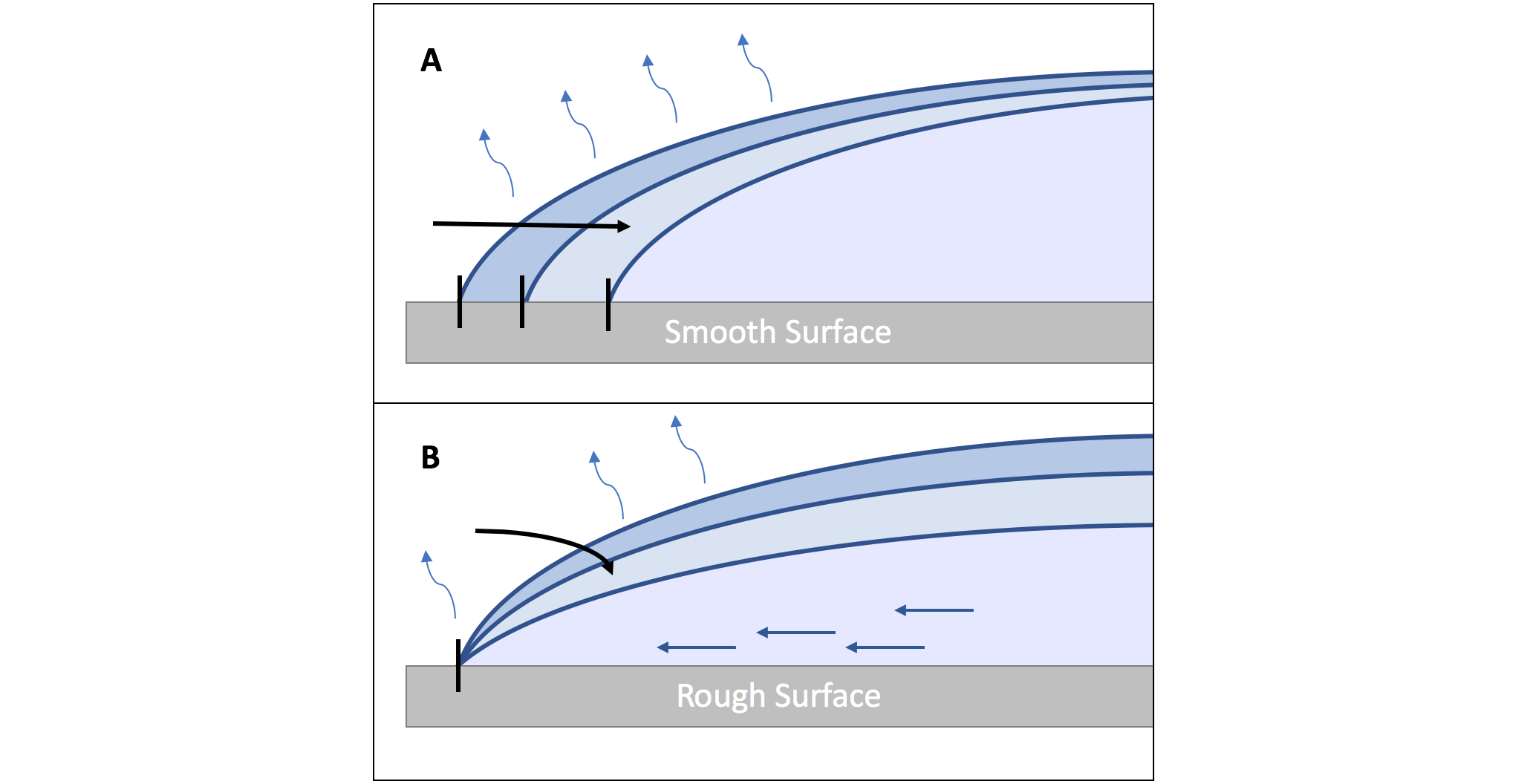
When sneezes are observed using high-speed photography, as in this video, an image of a turbulent cloud of droplets is revealed. These droplets break apart, collide, stretch and break apart again in a chaotic cascade that could exert considerable destructive shear forces on the viruses, but it is currently unknown whether the turbulence decreases the active virus concentration or if the virus particles influence the droplet break-up events. Solving this unknown may change the way we understand the role of air flows from ventilation systems in inhibiting active viruses.

Despite the lack of information on virus viability in the turbulent sneeze cloud, Poon and colleagues point out that studies suggest that the viral load of virus-carrying droplets is small – around 1 viable virus particle per droplet. However, sneezes typically generate up to 40,000 droplets, whereas a single cough (or talking for 5 minutes) will generate up to 3000. One also needs to know how many viruses a person must absorb before their dose becomes infectious – the minimum infective dose (MID). For the flu, the MID is reported to be around a few thousand, but the equivalent figure is not known for coronaviruses. All of these numbers play a role in infection but collectively point to the importance of limiting the amount of direct droplet transmission, and the importance of ventilation.

Finally, an airborne droplet containing a virus evaporates, at a rate depending on the local humidity, and will eventually completely dry. Interestingly, studies suggest that bacteriophages (viruses that invade bacteria) are more likely to survive in very dry or very humid air, but not in between. The reason for this seems to lie in how dissolved salt destructively interacts with the virus at steady (not slow or fast) evaporation rates. Other saliva components (like the gelation of the mucin proteins that make mucus slimy) also impact evaporation, further complicating the problem of viral transport and survival, but these clues indicate that controlling air humidity could be a viable option for hampering the virus.

On Surfaces

In the previous section, we’ve seen that how droplets behave in the air seems to be important, and this has been well-studied in a variety of commercial and academic contexts. As in ink-jet printing, we should also consider how droplets impact surfaces, as contact from surface to skin to mucus-membrane is another route through which viruses spread.

Here we will summarize the five main questions highlighted by Poon and colleagues in how droplets interact with surfaces, which could be important for understanding viral survival and transmission:

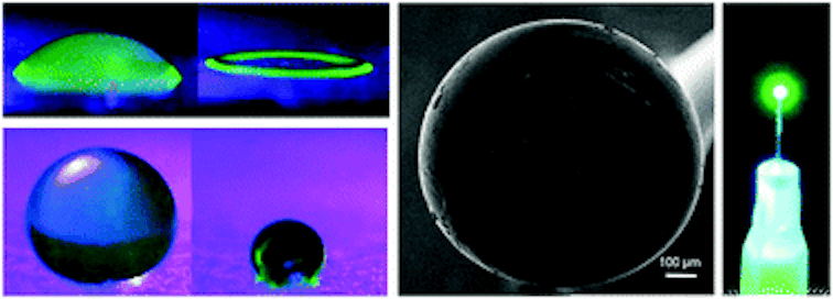
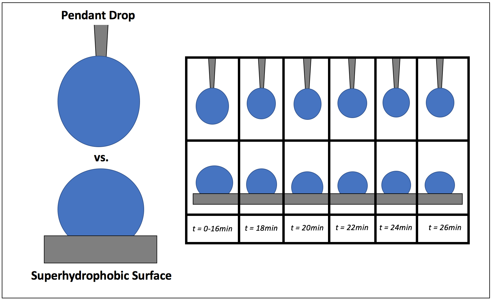
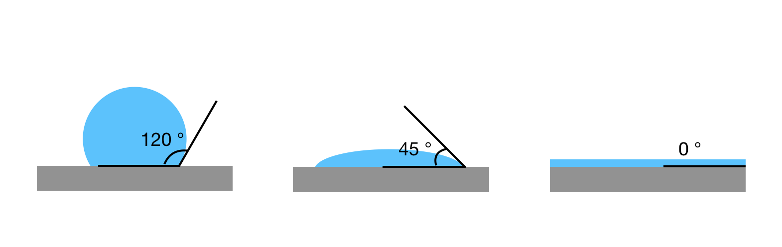
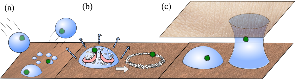
  • Splashdown. While ink-jet printed droplets need to avoid bouncing off surfaces, in a pandemic we ideally want the opposite. Polymers are known to give an anti-bounce property to droplets, but do mucins in saliva also have this effect? (see Figure 2 a)
  • Coffee rings. Much research has been done on the coffee-ring effect (When Espresso Evaporates), where suspended material is dragged to a droplet’s edges as it dries. Do viruses cluster at the edges in this way, and do these dragging forces impact their viability? This is further complicated by the salts and mucins present in these droplets, which will also accumulate at the edges (see Figure 2 b).
  • Material. Surfaces can have complex structures (smooth, ridged, or fibrous) and variable chemistry (metal, glass, or oily skin cells). Interestingly, virus viability tends to follow an exponential decay once on a surface, but the decay rate is higher on some surfaces (e.g. copper) than others (e.g. plastics). A question remains: is this due to a chemical catalytic property, or are some surfaces retaining moisture better than others?
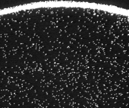
  • Capillary Bridges. Watery bridges can form when two wet surfaces come into close contact, but do microscopic bridges form when your skin comes into contact with surfaces, and do they enhance viral transport? Interestingly, bacteriophages are known to better transfer from surface to surface in humid environments, which suggests that such liquid bridges may be important (see Figure 2 c).
  • Fluidic Forces. It is well documented that bubbling gas through a solution or filtering it through a bed of glass beads deactivates viruses, but why? Poon argues that there could be significant capillary forces (which are known to deform latex particles in drying paint) pulling on virus particles if they get stuck at the interface between air and water, which will likely happen often in a vigorously bubbled or filtered solution. The behaviour of colloidal particles at interfaces is a rich area of ongoing study, so answers here are anticipated soon.
Figure 2. Schematic of processes to consider with viral loaded droplets on surfaces. (a). Droplets impacting surfaces. Bottom: droplet impact is absorbed, and it comes to rest in a single spherical cap. Middle: Droplet splashes into multiple droplets. Top: Droplet bounces away. (b) Droplet evaporation. Particles, salts, mucins and viruses accumulate at droplet edges via the coffee-ring effect. (c) Capillary bridges. Schematic shows a pathway viruses may take via capillary bridges between the resting surface and skin.

Final Note

The possibility of a global viral pandemic had been predicted by scientists for decades, and yet was not prevented, and the current one is seemingly unlikely to fully disappear in the near future. While the world’s hopes currently rest with developing and deploying effective vaccines, we should in parallel use every single weapon at our disposal to understand and treat the causes and symptoms of viral pandemics. The lessons we learn may also allow us to improve on some of the problems that the current measures are creating, such as the unfortunate and unsustainable use of single-use plastics in disposable protective gear that contribute to the “plastic pandemic”. Soft matter science may not be central to solving the problem of COVID-19, but is important, and could have a significant role to play in preventing the next global pandemic.

Featured image for the article is an edited version of a figure from the original article.

Self-assembling silk lasers

Rings, spheres, and optical resonators self-assembled out of silk

Original paper: 3D coffee stains


When I first learned about the coffee ring effect I thought it was super cool, but it seemed like an open-and-shut case. Why do rings form where some liquids, like spilled coffee, are left to dry? Roughness on the table causes the liquid to spread imperfectly across the surface, pinning the edges of the droplet in place with a fixed diameter. Because the diameter of the droplet can’t change during evaporation, new liquid must flow from the droplet’s center to the edges. This flow also pushes dissolved coffee particles to the edges of the droplet, where they are left behind to form a ring as the water evaporates away (Figure 1). More details can be found in our previous post, here. It’s a complicated phenomenon, but after being described in 1997 it doesn’t seem like anything new would be going on here. Right? Well, as usually happens in science, classic concepts have a way of popping back up in unexpected ways. Last year It?r Bak?? Do?ru and her colleagues in Prof. Nizamo?lu’s group at Koç University, Turkey published a study using the often troublesome coffee ring effect to their advantage: making self-assembling silk lasers.

pinning
Figure 1: Pinning and the Coffee Ring Effect. A cross section of a water droplet drying on a smooth surface (A) versus a rough surface (B). On a smooth surface the droplet shrinks due to evaporation. On a rough surface the edge of the droplet is pinned and cannot shrink, forcing an internal flow to maintain the droplet’s shape.

The fundamentals here are the same as the classic coffee ring effect, but instead of coffee particles Do?ru’s droplets hold a colloidal suspension of silk fibroin proteins. In a colloidal suspension, particles (such as proteins) are mixed in another material (such as water) and neither dissolve fully into solution nor precipitate out. Smoke, milk, and jelly are all examples of colloids. Harnessing the coffee ring effect to build 2D structures out of colloidal particles has been well developed since Witten’s description of the coffee ring effect in 1997 [1], but 3D self-assembly is much less common. What makes Do?ru’s 3D structures possible is the fibroin protein.

Fibroin is the primary component of silk from the silkworm Bombyx mori. These fibers have been used by humans for thousands of years to make textiles, but recently the fibroin protein has taken on new life when extracted from silk as an aqueous, water-based, suspension and regenerated into other forms [2,3]. Fibroin proteins are long, and they easily tangle up and bond to each other to form networks of layered crystalline structures called beta-sheets (?-sheets) (Figure 2). These sheets give silk fibers and other fibroin materials strength and toughness. Furthermore, fibroin materials are biocompatible and biodegradable.

Silk Fibroin and Beta Sheets
Figure 2: Silk Fibroin And ?-sheets. Silk is made of long fibroin proteins (a) that have a repeating molecular structure. These proteins bond together into ?-sheets (b), which then stack together (c) to form materials with high strength and toughness.

To create 3D structures with the coffee ring effect, Do?ru, Nizamo?lu, and their coworkers put droplets of silk solution on superhydrophobic surfaces. Superhydrophobic surfaces strongly repel water, preventing water-based liquids from spreading flat across the surface and making the droplets stand straight up during the drying process. This makes the angle between the edge of the droplet and the surface (called the contact angle) particularly high, between 95-145 degrees throughout evaporation. The interaction between water and the superhydrophobic surface determines the shape of the final structure, with high contact angles creating more spherical droplets (Figure 3). After a solid 2D ring of fibroin forms on the bottom, the silk proteins continue to stack along the droplet’s surface, forming a stable spherical shell of ?-sheets that the remaining water can evaporate through. The researchers found that the concentration of the fibroin solution was important for controlling the final structure. If the solution is too dilute then the shell will collapse in on itself, but if the fibroin concentration is too high the initial contact angle will be lower and the final structure will also be more 2D than 3D.

Contact Angle
Figure 3: Contact Angle. Droplets of the same solution show different contact angles on different surfaces (as adapted from Do?ru’s paper). On the left is a mildly hydrophobic surface, and on the right is a superhydrophobic surface. Note how the size of the contact angle (shown in white) increases with the hydrophobicity of the surface.

To make 3D spheres, the researchers tried the pendant drop method, hanging a droplet from the tip of a needle. Similar to getting high contact angles from a droplet on a hydrophobic surface, hanging a droplet from a needle gives that droplet a small contact area, and a spherical shape (Figure 4). If the diameter of the needle is the same size or smaller than the contact area of the droplet on a superhydrophobic surface, then the shape of a droplet squeezed out of the needle should be as or more spherical than the droplets in the previous experiment. In this study, the pendant drop method ends up producing more uniform drying. These pendant-drop shells are smooth enough inside to act as optical resonators, surfaces that reflect light waves back on themselves so the waves amplify each other (the “a” in “laser,” which I always forget comes from the acronym for “light amplification by stimulated emission of radiation”).

As a proof of concept, the researchers made shells out of fibroin mixed with green fluorescent protein (GFP). Fibroin ?-sheet formation is so stable that it still happens when small amounts of other materials are present, so the optical resonator can form in the same way it did with a fibroin-only solution. In this case, because GFP has been added, when the structure is exposed to the right light source it will amplify green light emitted by the shell itself – an “all protein laser” in the making.

Benefits of the Hanging Pendant Drop
Figure 4: Benefits of the Hanging Pendant Drop. The hanging pendant drop method can produce similar spherical drops to a hydrophobic surface. It was shown that the pendant drop method produces more spherical final structures (adapted from Do?ru’s paper).

Part of what’s exciting about this publication is that the authors harness the coffee ring effect for a fun new type of small scale, self-directed 3D “printing.” They showed that the method works for other polymers as well, but I agree with their choice to highlight the silk protein fibroin. Not only is fibroin biocompatible, but it also has the potential to be more environmentally friendly to process than other polymers and is already produced in large quantities globally as part of the textile industry.


[1] Han, W. and Lin, Z. “Learning from ‘Coffee Rings’: Ordered Structures Enabled by Controlled Evaporative Self-Assembly.” Angew. Chem. Int. Ed. 51 (2012): 1534–1546.

[2] Altman, G.H. et al. “Silk-based biomaterials.” Biomaterials 24 (2003): 401–416.

[3] Koh, L.-D. et al. “Structures, mechanical properties and applications of silk fibroin materials.” Prog. Polym. Sci. 46 (2015): 86–110.

When espresso evaporates: the physics of coffee rings

Original paper: Capillary flow as the cause of ring stains from dried liquid drops


fig1a
Figure 1. A 2-cm dried drop of coffee with a stain around the perimeter, forming a coffee ring. Adapted from Deegan et. al.

I’ve spilled a lot of coffee over the years. Usually not a whole cup, just a drop or two while pouring. And when it’s just a drop, it’s easy to justify waiting to clean it up. When the drop dries on the table, it forms a stain with a ring around the edges (Figure 1), giving it the look of a deliberately outlined splotch of brown in a contemporary art piece (when I say “coffee ring” I mean the small-scale, spontaneously formed stain around the edge of the original drop, rather than the imprint left on a table from the bottom of a wet coffee cup). But the appearance of these stains is simply a result of the physics happening inside the drop. Coffee is made of tiny granules of ground up coffee beans suspended in water, so the ring must mean that these granules migrate to the edge of the droplet when it dries. Why do the granules travel as they dry? Today’s paper by Robert D. Deegan, Olgica Bakajin, Todd F. Dupont, Greb Huber, Sidney R. Nagel, and Thomas A. Witten provides evidence that coffee rings arise due to capillary flow–the flow of liquid due to intermolecular forces within the liquid and between the liquid and its surrounding surfaces.

contact angle
Figure 2. Diagrams of contact angles for different droplets. From left to right, the first is exhibits poor wetting, with a large contact angle. The next has good wetting, with a smaller contact angle. The last has perfect wetting, with a contact angle of zero, and coffee grains suspended in this solvent would not be able to form a ring upon drying.

The researchers found that these rings don’t just form in coffee. Their observations showed that the rings form in a wide variety of solutes (the suspended coffee granules), solvents (the water), and substrates (the table you spill on) as long as a few conditions are met. First of all, the droplet has to have a non-zero contact angle[1] (See Figure 2). In other words, the droplet doesn’t spread out into a completely flat puddle on the table. Second, the contact line has to be pinned. This means that the surface has irregularities or roughness that cause the edge of the droplet to get stuck in place. Last, the solvent has to evaporate; the ring won’t form if the droplet never dries.

So now we know the conditions required for rings to form, but we want to know how they form. Deegan and his colleagues found that the rings are caused by a geometrical constraint. Here’s how it works: The pinning of the contact line means that the perimeter of the droplet cannot move, so the diameter of the droplet has to remain constant. But if the water in the droplet is evaporating, the droplet’s height will be reduced at every point (Figure 3a). Along the edges, where the droplet is thinnest, the height would be reduced to zero, and the droplet would shrink.

But the contact line pinning means that droplet can’t shrink. To prevent this shrinkage, liquid must flow out to replenish the liquid at the droplet edge as it evaporates. This flow brings with it the suspended coffee granules (or whichever solute is suspended in the solvent), pushing them outward until they pack at the edge of the droplet to form a ring (Figure 3b).

droplet cross sections
Figure 3. (a) Diagram showing the cross-section of a droplet on a surface. The shaded region shows how the droplet will shrink due to evaporation after a small amount of time if the contact line is not pinned. (b) Now, a black line is added to show how the droplet will shrink if the contact line is pinned. The arrows indicate that more liquid must flow to the outside of the droplet to replace what is lost to evaporation. Adapted from Deegan et. al.

By calculating how quickly water evaporates from the surface of a droplet, the researchers derived an expression for the mass of the ring as a function of time. It takes the form of a power law, which can be shown as a straight line on a log-log plot. Equipped with a quantitative prediction, the researchers set about performing experiments to test their model. Instead of using coffee, they opted for plastic microspheres suspended in drops of water. They placed the drops on glass slides and used a video microscope to image the droplets as they dried, recording the particles moving to the edges of the droplet (Figure 4).

video
Figure 4. Particles flowing to the edge of a droplet during evaporation to form a ring. Video from [2] and produced by Deegan et. al.
The researchers knew the mass of the individual particles, so they were able to calculate the mass of the ring as a function of time by counting the particles as they traveled to the edges. The results were shifted by an offset time t0 to account for early times where the power law prediction doesn’t hold and were shifted by mass M0 to account for the particles deposited during this initial stage. From the plot comparing the data and theory (Figure 5), we can see that the prediction shows good agreement with the data.

M vs T
Figure 5. Plot of mass in the ring as a function of time. The mass is plotted in units of particle number, so the plot shows how the number of particles grows over time. The three lines correspond to three different droplets. The upper curve overlapped with the middle so was shifted up for clarity. The circles show data and the solid lines show the theoretical prediction. The slope of 1.37 is the exponent of the power law predicted by the theory; On a log-log plot, a power law is a line with the exponent as the slope. Adapted from Deegan et. al.

In the twenty years since this paper was published, the study of drying droplets has continued in full force [3]. Scientists have discovered various particle patterns that can form under different drying conditions. Why do we care so much about these drying droplets? If the beauty of the physics isn’t motivation enough, then maybe the applications will convince you. The physics of drying is essential to inkjet printing, and a better understanding of the drying process could help make more precise printers [4]. Drying patterns can be used to identify the presence of certain proteins, making this a potential tool for disease detection [5]. Maybe next time you spill some coffee, you’ll take a moment to think of the physics of the drying droplet before you wipe it away.


[1] The contact angle is the angle where a liquid-gas interface meets a solid surface. The smaller the contact angle, the better the wetting of the surface.

[2] https://mrsec.uchicago.edu/research/highlights/coffee-ring-effect

[3] https://www.nature.com/uidfinder/10.1038/550466a

[4] Soltman, D. & Subramanian, V. Langmuir 24, 2224–2231 (2008).

[5] Trantum, J. R., Wright, D. W. & Haselton, F. R. Langmuir 28, 2187–2193 (2012)