Evolution usually solves challenges differently than human engineers—something easy for biology is often difficult for us, and vice versa. Learning from biology can help us solve difficult challenges more easily. One example of this is making complex optical lenses.
Figure 1. Lenses use refraction and geometry to direct light. (Figure by the author)
When light enters a new material it refracts, changing velocity and direction. A lens uses geometry and material properties to direct light on a specific path. You can see in Figure 1 how the shape and refractive index of a biconvex lens combine to direct light at a single spot. In biology, complex eyes like those found in most vertebrates and in squid have a lens that directs light onto the retina at the back of the eye, forming an image to be processed by the brain. Squid use spherical lenses to do this, but spherical lenses have a problem. As you can see in Figure 2, if you make a spherical lens out of one material (like glass), the light rays overlap after exiting the lens and the resulting image is blurry. This is called “spherical aberration.” Human engineers use spherical lenses a lot, and we correct for spherical aberration by combining multiple lenses. Squid, on the other hand, have evolved a lens that self-corrects for this distortion.
Figure 2. Spherical lenses usually produce blurry images, but not in squid. (Figure by the author)
We know, in theory, how a squid might do this. In 1854, the famous physicist James Clerk Maxwell mathematically designed a spherical lens with “perfect” focus. He showed that if the density of the lens changes along the radius, forming a density gradient that he called a “perfect medium,” then the lens will produce a clear image. Today engineers can make gradient index lenses like this, but the process is difficult and energy intensive. Squid evolved to grow them easily. Could understanding how squid make these lenses help human engineers learn to do the same thing? This question inspired Dr. Jing Cai and Prof. Alison Sweeney to study the structure of the squid lens.
You can see in Figure 3 that when you crack open the lens in a squid eye you find rings like the inside of a tree trunk. If each of these rings has a slightly different density, they could combine to create a perfect medium. The lens of an eye is made out of proteins called crystallins, which fold into individual particles before linking together into a single material. Cai and her collaborators discovered that the lenses of the Longfin inshore squid (Doryteuthis pealeii) use 53 different crystallin proteins of different sizes. They also found that the different proteins are used in different parts of the lens, and each layer of the lens has a slightly different structure. As you can see in Figure 4, small proteins at the center of the lens are densely packed together so that each protein is connected to six other proteins. However, the larger proteins at the edge of the lens have more space between them, and each protein only touches two others.
Figure 4. Protein particles assemble differently in different parts of the lens, creating a “perfect medium.” (Figure by the author)
This makes sense when you think about the cells that make these proteins. Cells rely on diffusion to bring building blocks to the right place for protein assembly and to send each assembled protein out to where it’s needed. When finished proteins link together to grow the lens, they disrupt this diffusion and stop protein production. By growing from dense to less dense and using so many different proteins (53 in the Longfin inshore squid), the cells are able to start and stop the growth of different layers while maintaining a single particle network. No part of the lens separates out or turns opaque, but there are still large enough regions with different densities to diffract light into alignment.
Cai and her collaborators showed that squid lenses definitely use a density gradient similar to Maxwell’s perfect medium to correct for spherical aberration. It’s likely that this density gradient not only creates a perfect medium, but also helps control lens assembly. Now that we know how squid build a perfect spherical lens, it is easier to envision how human engineers could grow our own complex optical materials.
The circulatory or vascular system of animals is a complex organ that performs multiple functions simultaneously. Take the human circulatory system for example: it transports oxygen and nutrients throughout the body, regulates temperature, helps in fighting diseases, etc. However, robots don’t usually function the same way. Despite the vast improvements in robot design, they still cannot do much multi-tasking. Part of the problem is that robots are typically built from rigid parts that only do one thing. A battery usually does not play any other role aside from providing energy, and moving parts are typically controlled by independent actuators. These single-purpose components (such as batteries) are typically less efficient than their biologic counterparts, and add to the weight of the robots (limiting their performance, maneuverability, speed, and autonomy).
In order to create multi-functional robots, we can take inspiration from biology and build them out of multi-purpose components. A research team led by Robert F. Shepherd from Cornell University (NY, USA) has found a way around this problem by integrating a multifunctional circulatory system into untethered, autonomous soft robots. In a recent issue of Nature, they present an aquatic soft robot with a synthetic circulatory system which provides chemical energy. The synthetic blood has zinc and iodide ions that make it electrically conducting (like an electrolytic blood), and powers an artificial heart (a pump that circulates “blood” throughout the robot body). Rather than using traditionally rigid materials to build the robot, the circulatory system is encased in a soft, flexible body made out of stretchable silicone. The circulating “blood” can deform the flexible body of the robot when pumped through the vascularized fins, like electrolytic “robot blood” flexing a muscle, moving the fins of the fish and propelling the robot forward (Figure 1, Video 1). By bringing materials and robot design closer to complex biological systems, their multifunctional synthetic vascular system can combine hydraulic force transmission, mechanical actuation, and energy storage (killing not two but three birds with one stone).
Figure 1. Lionfish-inspired robot powered by an electrolytic vascular system. a) Schematic shows synthetic blood in the tail fin (red) and in the dorsal and pectoral fins (yellow). b) The synthetic blood circulates through the robot body and actuates the tail fin by expanding and contracting the silicone parts at each side (adapted from Aubin et al.)
Although flow batteries are less energy dense than their lithium ion equivalents, their integration in soft robotic platforms has significant advantages: the electrolyte can fill up most of the volume of the robot and act as the hydraulic system (without the need of additional space), and the surface area of the flow batteries inside the soft robot can be maximized for an increased energy capacity (hence the large area of dorsal fins). With this approach, the autonomous robot can swim upstream for long operation times (up to 36 hours).
Video 1. Autonomous lionfish-inspired robot swimming.
The proposed bio-inspired approach reduces the gap between biological complex systems and synthetic, traditionally bulky robotic platforms. According to the authors, “this concept can be generalized to other machines and robots”, which opens a wide design space for multifunctional soft machines, from materials design to actuation and control. Such improvements in energy storage and performance could advance the development of soft robots for search-and-rescue operations, marine exploration, inspection of underwater pipelines, and coral reef health monitoring (all applications where autonomy and safety are critical). Although still in its infancy, “robot blood” could one day power, actuate, and control untethered soft robots that can safely and autonomously interact with humans in delicate environments. And just like in science fiction, robots with synthetic organs and circulatory systems could one day live among us.
The skeleton is the backbone of the body, both literally and figuratively. Healthy bones protect soft organs from injury and enable the body to move. Starting from childhood, staying active and following a healthy diet helps the body maintain healthy bones. However, as people age, their bones can start to weaken. There are often no early symptoms to weakening bones, and as a result the first indication of a problem may be a painful break once the weakening has already significantly progressed.
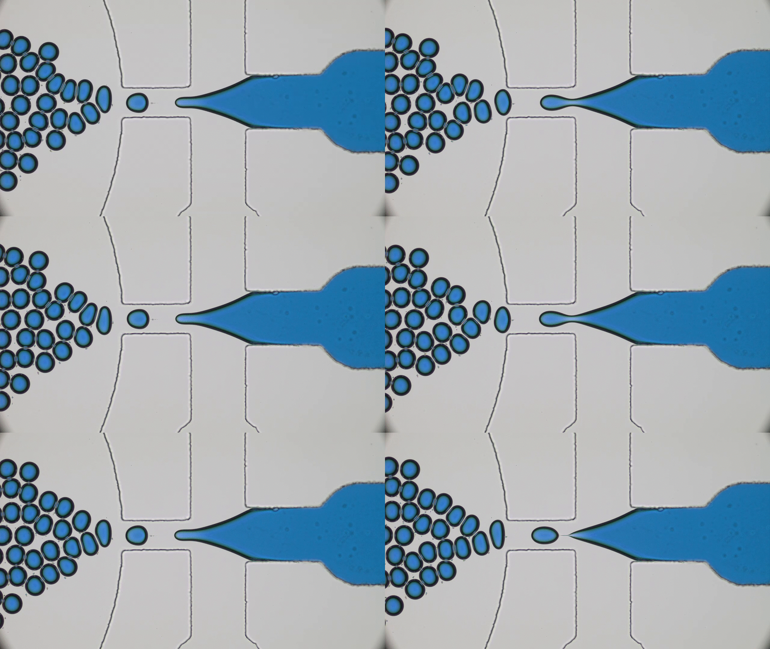
Although it may seem like bones are made of a hard material, they are actually an elegant combination of hard and soft materials. A primary component of bone is collagen, which forms a soft protein network. This network then provides a scaffold for calcium phosphate, a mineral that provides bone with its hardness and strength. This mixture of hard and soft material enables bone to be flexible enough to withstand impacts, but rigid enough to maintain its structural integrity.
Bone tissue, like other tissue in the body, is alive and therefore able to grow and heal. The material in bones is always being resorbed (or removed) by the body and simultaneously replaced. Around the age of thirty, the rate of resorption overtakes the rate of replacement, causing bones to slowly weaken. When these rates become too disparate, osteoporosis can develop, causing bones to become weak and porous [1].
Bone fractures are more common in patients with osteoporosis, and when they occur, treatment is often needed at the site of the break to promote bone regeneration. The typical treatment in these cases is an autologous bone graft, which involves taking bone from another area in the patient’s body and transferring it to the fracture site to help promote bone regrowth. This technique has two clear downsides: there is limited availability of bone tissue for grafting since it has to come from somewhere else in the person’s body, and removing bone tissue for a bone graft can cause damage to the donor site.
As a result, researchers are working to develop synthetic materials that can provide a better alternative to autologous bone grafts. A promising material would promote bone growth and be strong enough to sustain bending and support weight. Additionally, it needs to be non-toxic and not cause an immune system reaction. Synthetic bone graft materials already exist, but they do not work as well as bone grafts taken from the patient’s own body because they are not as mechanically strong and they do not promote as much bone growth. Hence, the search for better synthetic bone graft materials continues.
In today’s paper, Mani Diba and co-workers investigate a new synthetic material for use in the regeneration of bone tissue in osteoporotic patients. The material in question is a colloidal gel, which is a disordered network structure made of microscale particles suspended in a liquid. This network allows the material to resist applied forces and behave like a solid, even though it may be mostly made up of liquid. Colloidal gels are different from chemically covalent bonded gels [2], like jelly or agar, because their building blocks are microscale particles instead of polymers. These particles bond to each other mostly because they are hydrophobic, or water-repellent, so they would prefer to be next to each other than surrounded by water. The bonds between the colloidal particles are reversible, meaning they can break and reform more easily than covalent bondsin polymer gels, which allows the colloidal gel network to be more adaptable and reform after being broken apart.
The behavior of the colloidal gels is similar to that of toothpaste, which acts like a fluid as it is being forced out of the tube, but once it stops being squeezed it becomes more solid again and doesn’t flow off your toothbrush. For a bone graft material, this means that the colloidal gel can behave as a liquid as it is being injected into a bone defect, and then harden as the network reforms once it’s in place. While this is not a requirement for bone graft materials, it does make the material easier to put in place at a bone defect site.
The researchers prepare a colloidal gel by mixing gelatin particles and glass particles in water (see Figure 1). This choice of particles mimics the structure of bone tissue by using gelatin- a soft material- with glass, which is hard and provides mechanical strength. In order to be a good replacement for a bone graft, the gel must satisfy two main requirements. First, it needs to be mechanically robust to serve as a load-bearing scaffold for bone growth. Second, it needs to be biocompatible, meaning that it should support the growth of new bones.
Figure 1: (a) A schematic showing the formation of a gel by mixing glass and gelatin particles (b) Electron microscopy images of colloidal gelatin (left) and glass (right) particles (adapted from Diba et al.)
The first set of experiments in this paper look at the mechanical properties of the colloidal gel by measuring its storage modulus, which characterizes how strong the gel is. The researchers find that increasing the ratio of glass to gelatin particles or increasing the total number of particles in the gel increases the storage modulus by a factor of more than 100, from about 0.1 kilopascal to tens of kilopascals. The gel is also able to recover its initial storage modulus after being broken apart by shearing, similar to how silly putty can recover its mechanical properties after being stretched. This indicates that the network is able to reform in the bone and become solid again, as expected.
After characterizing the gel’s mechanical properties, the researchers investigate whether it can promote new bone growth. The growth of new bone starts with the multiplication of osteoblasts, or bone-forming cells, that produce bone matrix material. A signature of this process is an increase in the levels of certain enzymes. Once the matrix is well formed, it undergoes mineralization, which is the deposition of inorganic material (calcium) onto an organic matrix (collagen). This process can be monitored by measuring the amount of calcium added to the area [3]. The researchers track these two indicators, enzyme levels and calcium deposition, to measure the biocompatibility of the gels.
Diba and coworkers study the biocompatibility of the gels both in test tubes and in living animals. In the test tubes, they only find significant mineralization at a glass to gelatin ratio of 0.5 (the highest investigated), which also corresponds to the largest peak in enzyme levels. For testing in animals, the researchers therefore opt for a composite gel with a glass to gelatin ratio of 0.5 and compare the bone growth to that with a single-component gel with no glass particles. The researchers implant these gels in bone defects in the femurs of osteoporotic rats and measure the amount of bone growth after 8 weeks.
Surprisingly, in the rats, the addition of glass particles to the gel did not increase the amount of bone mineralization beyond that seen in the single-component gel as the researchers hoped. However, the bone growth in the composite gel did show more blood vessel-like structures than in the single component gels (see Figure 2), which is important because bone—like other living tissues—needs blood flow to supply oxygen and nutrients, as well as to remove waste products.
Figure 2: Images of bone regrowth from the composite gel in a rat femur. Left: Bone regrowth in a defect that was originally the size of the black circle. Center: Higher magnification image of the small green rectangle on the left. Black arrows point to blood vessel-like structures. Right: Higher magnification of the red rectangle in the center. Red arrows point to cells observed in the center of the original defect. (Adapted from Diba et al.)
Though the researchers in this study did not find the desired increase of bone mineralization in live rats by using a composite gel instead of a single-component gel, they did see other indicators of improvement. Including glass particles increased the storage modulus of the gel, indicating more mechanical strength. They also saw indicators of improved biocompatibility. The bone growth in the composite gel showed an increase in blood vessel-like structures, and they found test tube results which suggested that including the glass particles may still improve mineralization if a higher ratio of glass to gelatin is used. Considering these improvements over a single-component colloidal gel, this composite colloidal gel is a promising development in the search for better bone graft materials.
[2] Covalent bonding is the sharing of valence electrons, which are in the outer shell of electrons, between atoms to make a full valence shell. Any time two non-metals come together they will share their valence electrons.